Model for simulating and demonstrating pathogenesis of pulmonary edema caused by mitral valve stenosis

A technology for mitral valve stenosis and pulmonary edema, applied in teaching models, educational appliances, instruments, etc., can solve the problems that students are difficult to understand the dynamic process, the content is complex and abstract, and the students are difficult to understand hemodynamic changes.

Inactive Publication Date: 2012-04-04
关真民
View PDF5 Cites 3 Cited by
  • Summary
  • Abstract
  • Description
  • Claims
  • Application Information

AI Technical Summary

Problems solved by technology

Due to the complex and abstract content, it is difficult for students to understand the dynamic process
In the experimental teaching, the pathological characteristics

Method used


Image

  • Model for simulating and demonstrating pathogenesis of pulmonary edema caused by mitral valve stenosis
  • Model for simulating and demonstrating pathogenesis of pulmonary edema caused by mitral valve stenosis

Examples


Embodiment Construction

[0021] A specific embodiment of the present invention provided by the accompanying drawings below will further describe its structure.

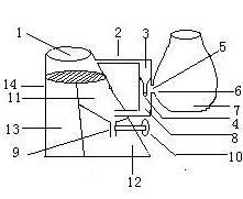
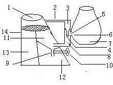
[0022] See attached figure 1 The conus pulmonary artery model 1 of the device is placed above the heart model 14 and connected to the pulmonary artery branch model 2 . The heart model 14 is divided into a right heart 13 , a left atrium 11 , and a left ventricle 12 . One end of the alveolar wall capillary model 3 is connected to the pulmonary artery branch model 2, and the other end is connected to the pulmonary vein model 8. The alveolar wall hole 6 corresponds to the alveolar wall capillary hole 5. The pulmonary vein model 8 enters the heart model left atrium 11, mitral valve The leaf regulating switch 10 regulates the opening and closing of the mitral valve 9 to regulate the blood flow from the left atrium 11 to the left ventricle 12 of the heart model.

[0023] [0008] When in use, add clear water to the conus pulmonary artery model 1, ...


PUM


Abstract

The invention relates to a model for simulating and demonstrating the pathogenesis of pulmonary edema caused by mitral valve stenosis, belonging to a medical teaching facility, in particular to a teaching tool for demonstrating the pathogenesis of pulmonary edema caused by mitral valve stenosis. The teaching tool mainly comprises a conical pulmonary artery model, a branched pulmonary artery model, an alveolar wall capillary model, an alveolar model, a pulmonary vein model, a heart model, a mitral valve model and a mitral valve cusp adjustment switch which form a model system for simulating and demonstrating the pathogenesis of pulmonary edema caused by mitral valve stenosis. The device has a simple structure, is convenient to operate, can be used to teach intuitively and pellucidly and is beneficial to culturing the problem observing and analyzing capacity of the students.

Description

technical field [0001] The invention belongs to medical teaching equipment, in particular to teaching aids for demonstrating the mechanism demonstration model of pulmonary edema caused by mitral valve stenosis. Background technique [0002] In the traditional teaching of the mechanism of pulmonary edema caused by mitral valve stenosis, the teaching method is mainly used: after mitral valve stenosis → blood flow from the left atrium into the left ventricle is blocked → blood stasis in the left atrium → pulmonary venous return is blocked → pulmonary congestion, Pulmonary edema, and then deduce the corresponding clinical manifestations, such as dyspnea, lip cyanosis, etc. [1] 。 Due to the complex and abstract content, it is difficult for students to understand the dynamic process. In the experimental teaching, the pathological characteristics of pulmonary congestion and pulmonary edema are mainly observed through pathological slices, and it is difficult for students to underst...

Claims


Application Information

Patent Timeline
IPC IPC(8): G09B23/28
Inventor 关真民鹿勇侯亚莉
Owner 关真民
Why Eureka
Try Eureka