
Image navigation-based parallel robot-assisted artificial cervical intervertebral disc replacement surgery positioning method
A technology of robot-assisted and positioning method, which is applied in the field of surgery with parallel robots, and can solve the problem of doctors being exposed to more radiation
- Summary
- Abstract
- Description
- Claims
- Application Information
AI Technical Summary
Problems solved by technology
Method used
Image

Examples
specific Embodiment approach 1
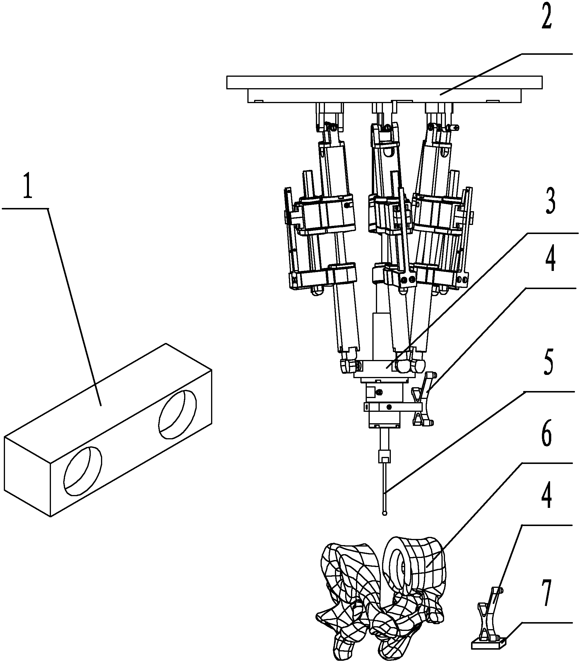
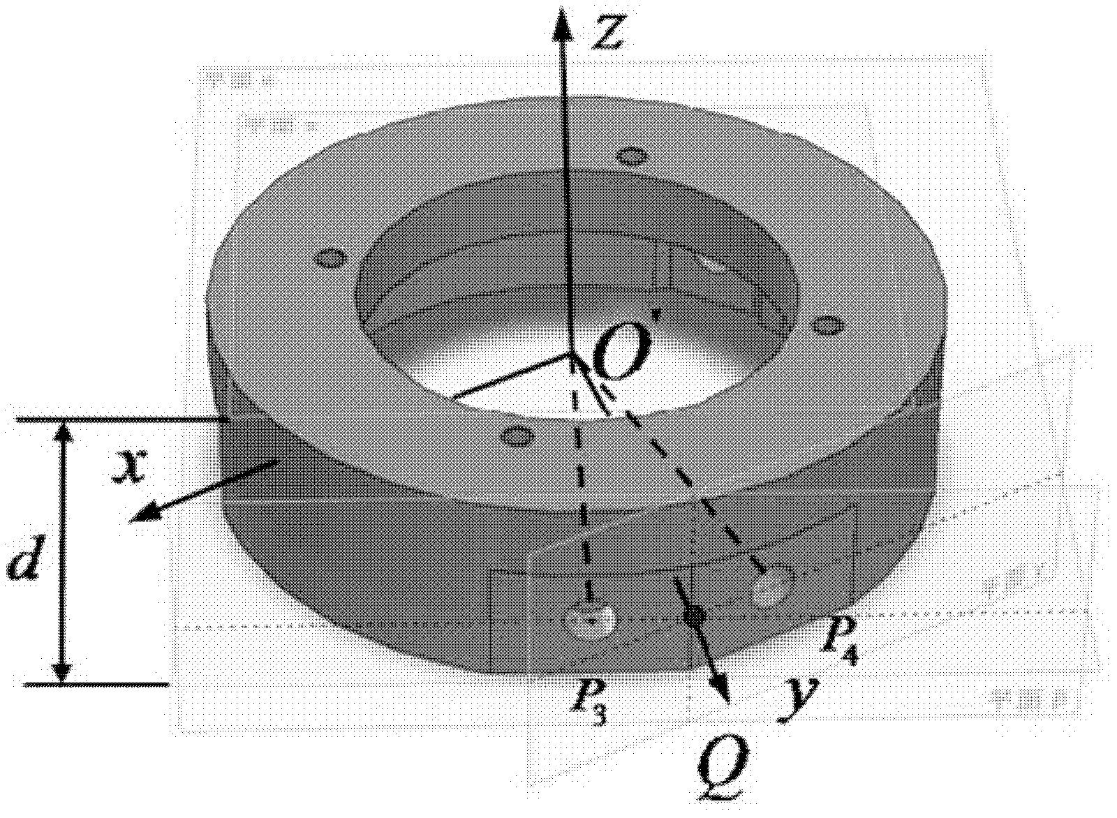
[0029] Specific implementation mode one: as Figure 1~5 As shown, the positioning method for parallel robot-assisted artificial cervical disc replacement surgery based on image navigation described in this embodiment is realized based on an optical positioning system, a parallel robot, a parallel robot grinder 5 and a computer, and the optical positioning system includes Two reflectors 4, an optical locator 1 and a probe, the parallel robot and the optical locator are connected to the computer, and a reflector 4 is installed on the drill shank of the parallel robot grinding drill 5; the specific method of the described method The process is:
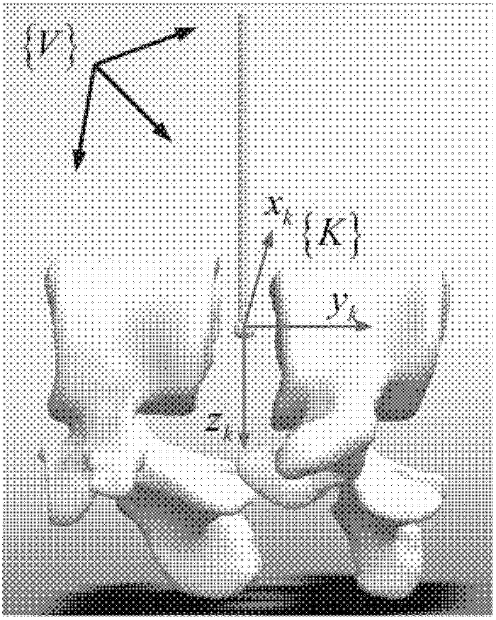
[0030]Step A, utilize the three-dimensional reconstruction software to complete the three-dimensional reconstruction of the cervical vertebra CT image at the patient's lesion by the computer, obtain the three-dimensional model of the cervical vertebra of the disease, and use the solidworks software to grind and drill (parallel robot grin...
PUM
 Abstract
Description
Claims
Application Information