
Radiographic system

A technology of radiography and radiation, applied in the fields of radiological diagnostic equipment, medical science, mammography, etc., which can solve problems such as easy movement, disease cannot remain still for a long time, contrast and resolution deterioration, etc.

Inactive Publication Date: 2012-07-11
FUJIFILM CORP
View PDF1 Cites 8 Cited by
  • Summary
  • Abstract
  • Description
  • Claims
  • Application Information

AI Technical Summary

Problems solved by technology

In particular, when multiple imaging is performed with respect to X-ray phase imaging, imaging should be performed in a short time because patients generally cannot remain still for a long time due to disease, and thus tend to move
Artifacts are produced in phase contrast images when the subject moves during imaging, with significant degradation in contrast and resolution

Method used


Image

  • Radiographic system
  • Radiographic system
  • Radiographic system

Examples


Embodiment Construction

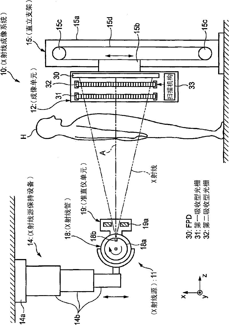
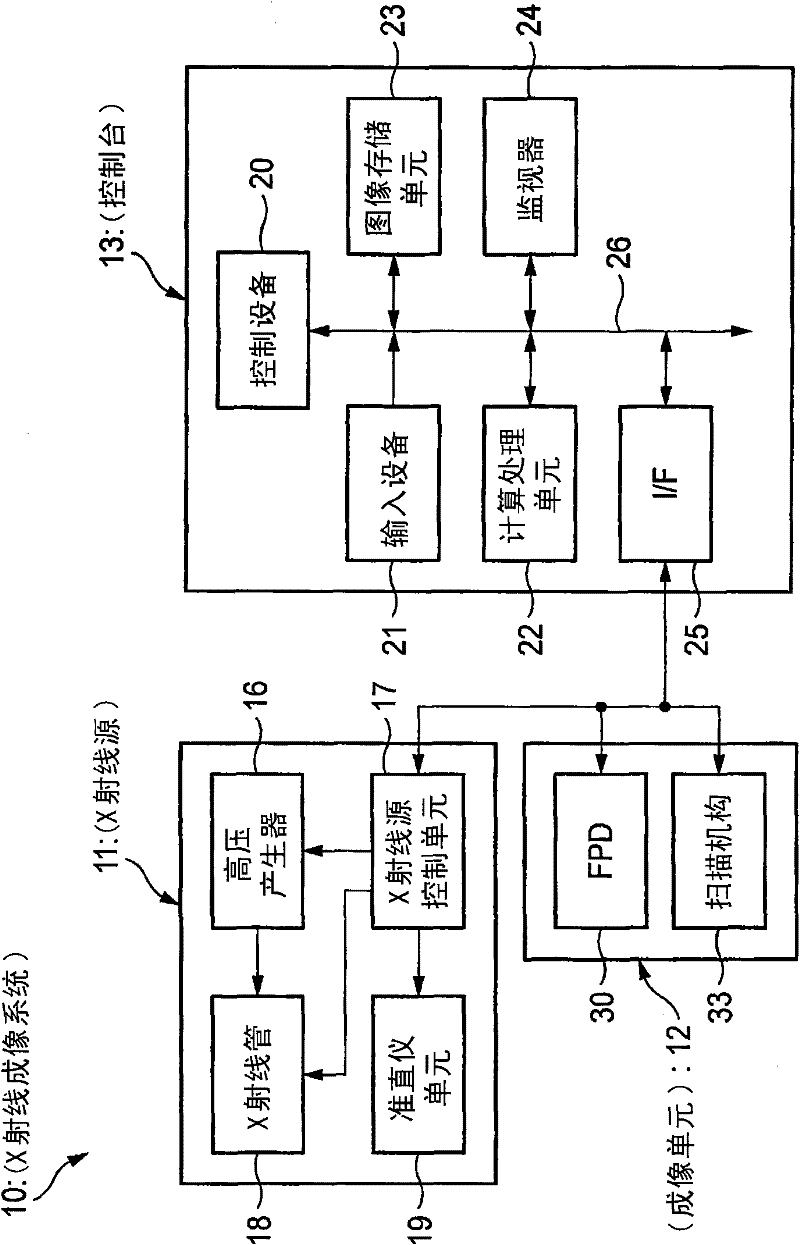
[0031] figure 1 An example of the configuration of a radiographic system for explaining an illustrative embodiment of the present invention is shown, and figure 2 Yes figure 1 Control block diagram of the radiography system.

[0032] The X-ray imaging system 10 is an X-ray diagnostic device that performs imaging on an irradiated object (patient) H while the patient is standing, and includes: an X-ray source 11 for irradiating the irradiated object H with X-rays; an imaging unit 12, and X-ray source 11 is opposite, detects the X-ray that has penetrated object H from X-ray source 11, and thus produces image data; And console 13, based on operator's operation, controls the exposure operation of X-ray source 11 and The imaging operation of the imaging unit 12 calculates image data acquired by the imaging unit 12 and thereby generates a phase contrast image.

[0033] The X-ray source 11 is held by the X-ray source holding device 14 suspended from the ceiling so that it can move...


PUM


Abstract

A radiographic system includes: a first grating; a second grating having a period that substantially coincides with a pattern period of a radiological image formed by radiation having passed through the first grating; a radiological image detector that detects the radiological image masked by the second grating and outputs image data of the detected radiological image, and a control unit that performs a switching between a first mode in which a plurality of imaging is performed with the second grating being positioned at relative positions having different phases with regard to the radiological image and a second mode in which the radiological image detector is driven without radiation exposure. The control unit repeatedly drives the radiological image detector in the second mode until the radiological image detector is in a steady state and shifts to the first mode after the radiological image detector is in the steady state.

Description

technical field [0001] The present invention relates to radiography systems. Background technique [0002] Since X-rays attenuate with the number of atoms of the elements that make up the substance and the density and thickness of the substance, they are used as probes to see inside the object being photographed. In the fields of medical diagnosis, non-destructive inspection, etc., X-rays are widely used for imaging. [0003] In a general X-ray imaging system, an irradiated object is arranged between an X-ray source that irradiates X-rays and an X-ray image detector that detects an X-ray image, and a transmission image of the irradiated object is captured. In this case, the X-rays irradiated from the X-ray source to the X-ray image detector are attenuated (absorbed), and then incident into the X-ray image detector, the attenuation (absorption) value depends on the Differences in material properties (eg, atomic number, density, and thickness) exist in the path of the image ...

Claims


Application Information

Patent Timeline
Patent Type & Authority Applications(China)
IPC IPC(8): A61B6/00
CPCA61B6/4233A61B6/502A61B6/4035A61B6/484A61B6/4291
Inventor 岩切直人
Owner FUJIFILM CORP
Learn More