
Magneto-acoustic microscopic imaging method and imaging system

A technology of microscopic imaging and imaging system, applied in the directions of acoustic wave diagnosis, infrasound wave diagnosis, material magnetic variables, etc., can solve the problems of difficult real-time imaging, long time, undiscovered literature and patent reports, etc.

Active Publication Date: 2012-11-21
INST OF ELECTRICAL ENG CHINESE ACAD OF SCI
View PDF5 Cites 38 Cited by
  • Summary
  • Abstract
  • Description
  • Claims
  • Application Information

AI Technical Summary

Problems solved by technology

[0006] It can be seen from the development process of magnetoacoustic imaging that the excitation frequency of the pulsed magnetic field is 1MHz, which fully meets the clinical application of future medical magnetoacoustic imaging, but as a microscope, this frequency is far from enough
In terms of reconstruction algorithms, the reconstruction algorithms of magnetoacoustic imaging are mainly filter back projection algorithm (Xu 2004), algorithm based on acoustic reciprocity theorem (Xia 2009), and reconstruction algorithm based on potential function (Xia 2010). In order to realize the realization, the sample must be scanned in a closed circle to obtain the sound pressure signal to realize the reconstruction of the image. Through the closed circle scan method, the sound pressure signal obtained at each detection point is transmitted from any point of the sound source in the detection section of the sample to the detection point. The signal superposition, that is, the detection signal at any point in the scanning period is the collection of sound pressure signals of all point sound sources in the excitation source area. The advantages of these traditional imaging algorithms are that they avoid the limitation of the diffraction effect of sound waves and can achieve high resolution. Imaging, but because the reconstruction algorithm needs to scan the object and average the data, it takes a long time and it is difficult to image in real time
[0007] Many research institutions at home and abroad have carried out research on magnetoacoustic imaging methods. However, the current research mainly focuses on how to realize the clinical application of magnetoacoustics. In terms of magnetoacoustic microscopic imaging, no relevant literature and patent reports have been found.

Method used


Image

  • Magneto-acoustic microscopic imaging method and imaging system
  • Magneto-acoustic microscopic imaging method and imaging system
  • Magneto-acoustic microscopic imaging method and imaging system

Examples


Embodiment Construction

[0019] The present invention will be further described below in conjunction with the accompanying drawings and specific embodiments.

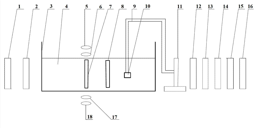
[0020] The imaging method of the present invention is based on the principle of magnetoacoustic electric imaging: pulse excitation 2 is applied to the conductive target imaging body 7 placed in the static magnetic field, and an induced eddy current is generated in the conductive target imaging body 7 . The combined action of the induced eddy current and the static magnetic field generates Lorentz force, which causes the vibration of the particle in the conductive target imaging body 7 and generates an ultrasonic signal. Using the imaging principle of the acoustic lens, the array ultrasonic probe 10 is used on the focal plane of the acoustic lens 8 to receive the image signals of the ultrasonic signals of each particle in the conductive target imaging body 7 . The received image signals of the ultrasonic signals of each particle are sequentially...


PUM


Abstract

The invention discloses a magneto-acoustic microscopic imaging method, comprising the steps of applying pulse excitation on a conductive target imaging body in a static magnetic field, generating an inductive vortex in the conductive target imaging body, and generating a Lorentz force by the interaction of the inductive vortex and the static magnetic field so as to cause the vibration of mass points in the imaging body to generate ultrasonic signals; receiving image signals of the ultrasonic signals of each mass point in the conductive target imaging body by using an array ultrasonic probe on a focal plane of an acoustic lens, imaging the received image signals of each mass point in the conductive target imaging body, so that each mass point image signal is proportional to the Lorentz force divergence of a corresponding point in the conductive target imaging body, and a Lorentz force divergence image of the conductive target imaging body or a reconstructed image according to current density rotation can be obtained according to the image signals of the ultrasonic signals detected by the array ultrasonic probe. A magneto-acoustic microscopic imaging system using the imaging method disclosed by the invention comprises a synchronous trigger and control module (1), an excitation source, an imaging system and a week signal detecting system.

Description

technical field [0001] The invention relates to an electrical impedance imaging method, in particular to a magnetoacoustic electrical impedance microscopic imaging method and device. Background technique [0002] Magneto-acoustic tomography (MAT) is a very promising medical imaging method that has emerged in recent years. It has the advantages of high contrast and high penetration. It uses static magnetic fields and pulsed magnetic fields to act on biological tissues to generate ultrasonic waves. , for resistivity reconstruction by measuring the ultrasound signal. As a novel imaging method, this method has many potential advantages in performing structural and functional microscopic imaging of biological tissues: [0003] (1) Magnetoacoustic signals depend on both the electrical properties of tissues and the acoustic properties of biological tissues. Compared with pure ultrasonic microscopy, magnetoacoustic microscopy provides more information and is a powerful supplement t...

Claims


Application Information

Patent Timeline
Patent Type & Authority Applications(China)
IPC IPC(8): G01N27/72A61B5/053A61B8/00
CPCA61B5/0093
Inventor 夏慧刘国强黄欣王霜陈晶
Owner INST OF ELECTRICAL ENG CHINESE ACAD OF SCI
Learn More