Method for measuring myocardium ultrasonic angiography image physiological parameters based on empirical mode decomposition (EMD)

A technology of empirical mode decomposition and contrast-enhanced ultrasound, applied in image data processing, image analysis, instruments, etc., can solve the problems of limited clinical practical value and inability to express the nature of the area to be diagnosed

Inactive Publication Date: 2013-01-02
HARBIN INST OF TECH
View PDF1 Cites 13 Cited by
  • Summary
  • Abstract
  • Description
  • Claims
  • Application Information

AI Technical Summary

Problems solved by technology

[0009] Clinically, fitting curves are often used to qualitatively measure the myocardial parameters of contrast medium perfusion, but such measurement methods are very dependent on the experience of doctors, and there are certain measurement errors; at the same time, since the fitting curve only reflects the time-average grayscale The change trend information of the curve, but more informat

Method used


Image

  • Method for measuring myocardium ultrasonic angiography image physiological parameters based on empirical mode decomposition (EMD)
  • Method for measuring myocardium ultrasonic angiography image physiological parameters based on empirical mode decomposition (EMD)
  • Method for measuring myocardium ultrasonic angiography image physiological parameters based on empirical mode decomposition (EMD)

Examples


specific Embodiment approach 1

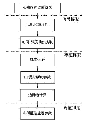
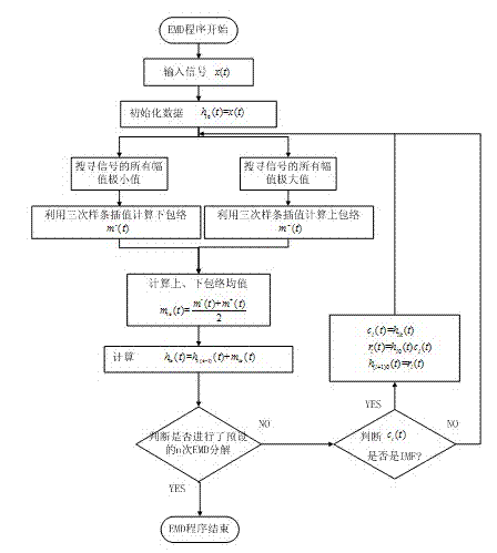
[0032] Specific implementation mode one: as figure 1 As shown, the method for measuring physiological parameters of myocardial contrast-enhanced ultrasound images based on empirical mode decomposition in this embodiment includes the following steps: firstly, the myocardial region of the myocardial contrast-enhanced image is divided into 6 regions in a clockwise direction, and each region is extracted respectively. The time-intensity curve in the process; use the EMD method to decompose the time-intensity curve to obtain the eigenmode functions corresponding to different frequencies; perform Hilbert transformation on the eigenmode functions respectively to obtain various instantaneous characteristic parameters, and finally get Hilbert marginal spectrum, according to the judgment of the energy threshold, can obtain the physiological parameters of myocardial perfusion corresponding to the myocardial contrast image, including the range of respiratory frequency and heartbeat frequen...

specific Embodiment approach 2

[0073] Specific implementation mode 2: In order to evaluate the time-intensity curve analysis of myocardial ultrasound images by using empirical mode decomposition, and the measurement and research of physiological parameters in the myocardial perfusion process by using marginal spectrum, this implementation mode takes the myocardial contrast-enhanced ultrasound images of SD rats as an example , to implement the above process in detail.

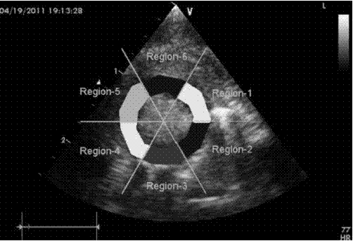
[0074] Execute Step 1: Carry out six-region segmentation on the myocardial contrast-enhanced ultrasound images of SD rats, such as image 3 Shown, represent the anterior wall myocardial area, lateral wall myocardial area, posterior wall myocardial area, inferior wall myocardial area, posterior septal myocardium area and anterior septal myocardium area of ​​rat myocardium, respectively.

[0075] The time-intensity curves of rat myocardial contrast-enhanced ultrasound images were extracted. Taking time as the abscissa (each ultrasound image fr...


PUM


Abstract

The invention relates a method for analyzing a time-intensity curve of a myocardium ultrasonic angiography image, in particular to a method for measuring myocardium ultrasonic angiography image physiological parameters based on EMD. The method includes dividing a myocardial area into six areas according to a circumference, extracting perfusion signals and extracting a time-intensity curve of each of divided myocardial areas respectively; performing EMD on an extracted time-average gray value intensity curve to obtain an expanded signal intrinsic mode function, performing Hilbert transform on the obtained first intrinsic mode function to obtain instantaneous spectrum parameters, and adding amplitude values of each frequency point to obtain a marginal spectrum; and performing threshold judgment on a marginal spectrum through energy thresholds of the marginal spectrum. According to the method, an analytical approach based on the myocardium ultrasonic angiography image is provided for diagnosing myocardial infarction or myocardial microcirculation, and further physiological parameter measurement accuracy based on the myocardium ultrasonic angiography image is improved, and doctor subjective dependence is reduced.

Description

technical field [0001] The invention relates to a method for analyzing time-intensity curves of myocardial ultrasound contrast images, in particular to a method for feature extraction of time-intensity curves by using empirical mode decomposition and a method for measuring physiological parameters by using Hilbert marginal spectrum. Background technique [0002] The heart is an important organ of the human body. With each contraction of the heart, the blood carrying oxygen and nutrients is transported to the whole body through the aorta to supply the metabolic needs of various tissue cells. Due to abnormal lipid metabolism, lipids in the blood deposit on the smooth coronary intima, forming thrombus. Once microthrombosis forms, it will cause severe stenosis or even blockage of microvessels, resulting in abnormal microcirculation perfusion. In severe cases, acute myocardial ischemia, necrosis, or even life-threatening. Even if coronary revascularization is used for treatment,...

Claims


Application Information

Patent Timeline
IPC IPC(8): G06T7/00
Inventor 金晶杜国庆沈毅
Owner HARBIN INST OF TECH
Why Eureka
Try Eureka