Rapid imaging method of blood vessel without contrast agent

An imaging method and contrast agent technology, applied in the field of medical imaging, can solve problems such as direct application of vascular imaging

Active Publication Date: 2013-04-24
SHANGHAI UNITED IMAGING HEALTHCARE
View PDF6 Cites 3 Cited by
  • Summary
  • Abstract
  • Description
  • Claims
  • Application Information

AI Technical Summary

Problems solved by technology

Since the position of the signal in the image domain obtained by N / 2 ghost imaging is inconsistent with the position of the signal source, the existing parallel accelerated imaging method in the image domain cannot be directly applied to N / 2 ghost imaging
[0007] The present invention overcomes the technical defect that the parallel accelerated imaging method in the prior art cannot be directly applied to blood vessel imaging of N / 2 ghost images, and proposes a fast imaging method of blood vessels without contrast agent, combining the improved parallel accelerated imaging method with N / 2 Ghost Imaging Combined and Applied to Contrast Agent-Free Vascular Imaging

Method used


Image

  • Rapid imaging method of blood vessel without contrast agent
  • Rapid imaging method of blood vessel without contrast agent
  • Rapid imaging method of blood vessel without contrast agent

Examples


Embodiment 1

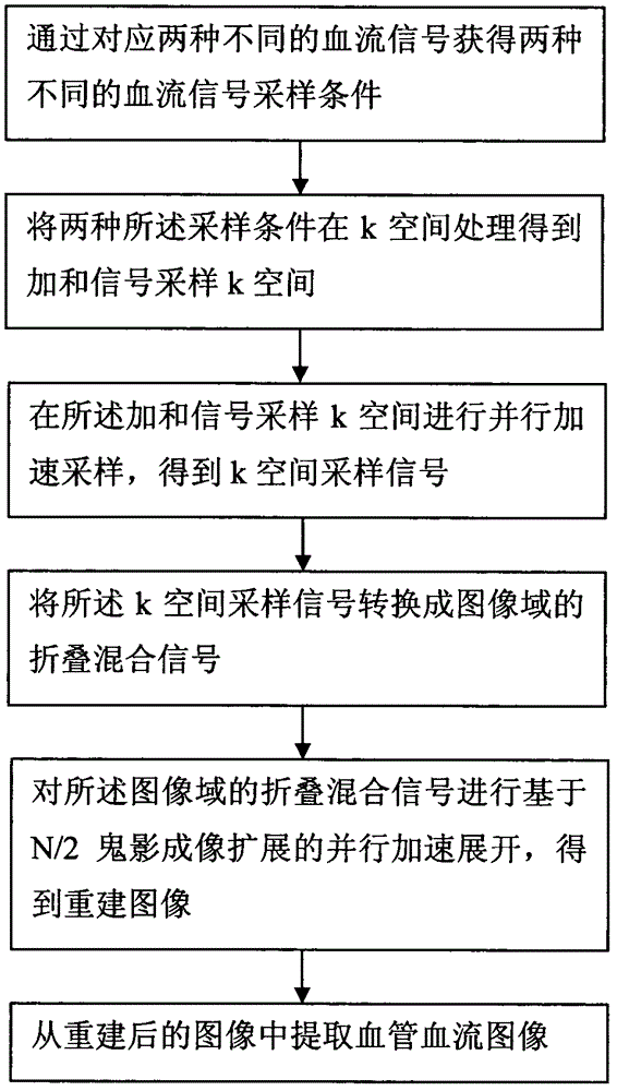
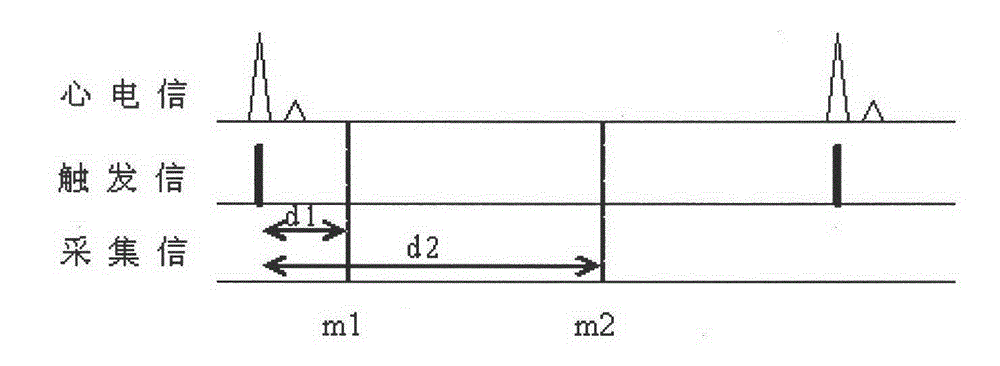
[0083] Such as image 3 As shown, by sampling the signals after two different delay times of ECG gating, for example, the delay times are d1=0.1 second and d2=0.5 seconds respectively, the sampling conditions m1 and m2 of two different blood flow states are determined. The magnetic resonance signals corresponding to the sampling conditions m1 and m2 are M1 and M2, respectively.

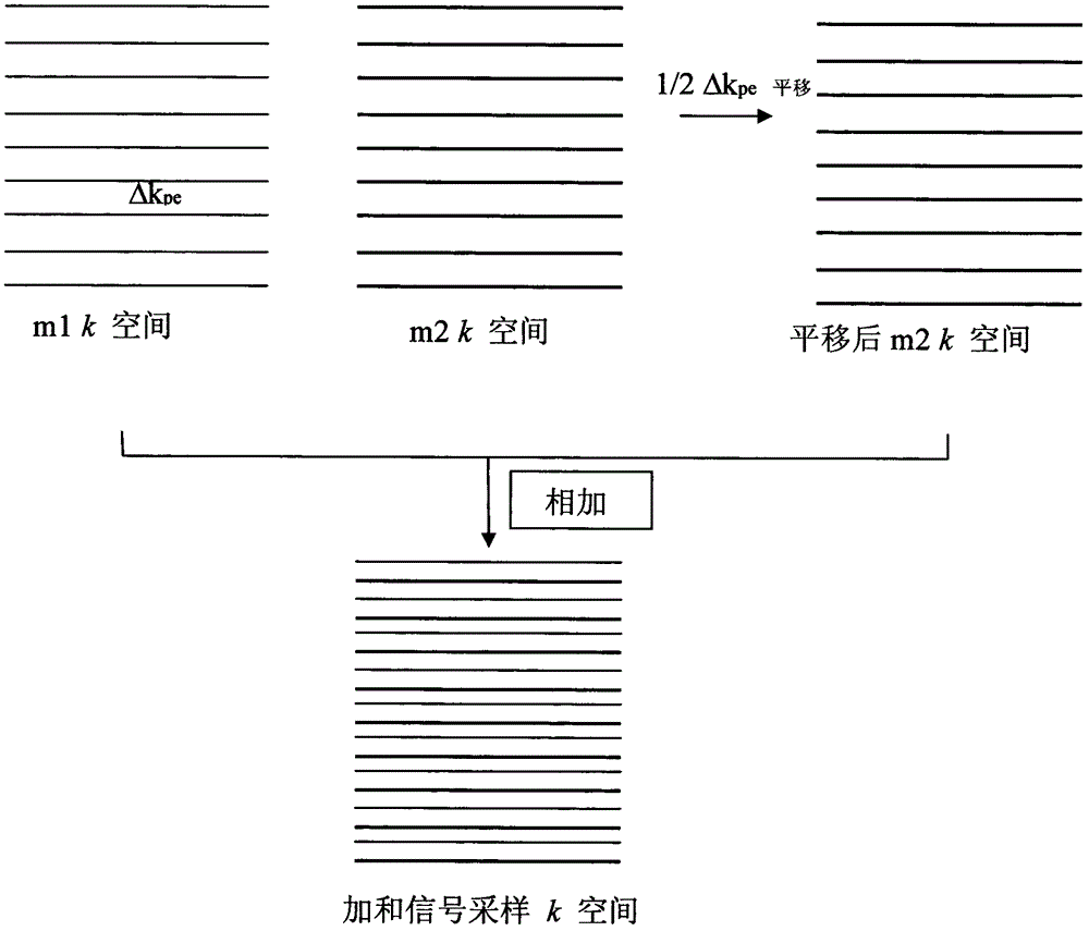
[0084] Referring to the N / 2 ghost imaging technology, the above two sampling conditions are alternately combined in k-space to obtain the sum signal sampling k-space, such as figure 2 As shown, the m2 k-space is shifted half a step along the direction of phase encoding, and then the shifted m2 k-space is merged with the m1 k-space to obtain the sum signal sampling k-space.

[0085] Parallel accelerated sampling in summed signal sampling k-space. For example, if the acceleration factor is R=3, when sampling the k-space of the summed signal, only the first of every three data lines is collected along ...

Embodiment 2

[0092] Such as Figure 4 As shown, under the same ECG gating condition, two different flow compensations are used in the imaging sequence, such as no flow compensation at m1 and flow compensation at m2, to determine the sampling conditions of two different blood flow states m1 and m2. The magnetic resonance signals corresponding to the sampling conditions m1 and m2 are M1 and M2 respectively.

[0093] Referring to the N / 2 ghost imaging technology, the above two sampling conditions are alternately combined in k-space to obtain the sum signal sampling k-space, such as figure 2 As shown, the m2 k-space is shifted half a step along the direction of phase encoding, and then the shifted m2 k-space is merged with the m1 k-space to obtain the sum signal sampling k-space.

[0094] Parallel accelerated sampling in summed signal sampling k-space. For example, if the acceleration factor is R=3, when sampling the k-space of the summed signal, only the first of every three data lines is ...

Embodiment 3

[0101] Such as Figure 5 As shown, by sampling the magnetic reversal of protons upstream or downstream of the imaging area, such as using a 180-degree adiabatic radio frequency pulse to achieve magnetic reversal, the sampling conditions m1 and m2 of two different blood flow states are determined. The magnetic resonance signals corresponding to the sampling conditions m1 and m2 are M1 and M2 respectively.

[0102] Referring to the N / 2 ghost imaging technology, the above two sampling conditions are alternately combined in k-space to obtain the sum signal sampling k-space, such as figure 2 As shown, the m2 k-space is shifted half a step along the direction of phase encoding, and then the shifted m2 k-space is merged with the m1 k-space to obtain the sum signal sampling k-space.

[0103] Parallel accelerated sampling in summed signal sampling k-space. For example, if the acceleration factor is 3, when sampling the k-space of the sum signal, only the first of every three data lin...


PUM


Abstract

The invention discloses a parallel accelerating imaging method of a blood vessel without contrast agent. The rapid imaging method of the blood vessel without contrast agent includes the following steps: obtaining two different blood flow signal sampling conditions by corresponding to two different blood fluid signals, processing the two different blood flow signal sampling conditions in a k space to obtain an additive signal sampling k space, conducing parallel acceleration sampling in the additive signal sampling k space to obtain k space sampling signals, transforming the k space sampling signals into folded and mixed signals in an image field, spreading the parallel acceleration which is based on N / 2 ghost imaging extension to obtain extended image field signals, and extracting blood flow images of the blood vessel from the extended image field signals. The rapid imaging method of the blood vessel without contrast agent improves the existing parallel acceleration imaging method, extends the corresponding relation of pixel position of various image areas and sensitivity of a radio-frequency coil and enables the improved parallel acceleration imaging method to be combined with the N / 2 ghost imaging method and to be applied to the blood flow imaging of the blood vessel without contrast agent.

Description

technical field [0001] The invention belongs to the field of medical imaging, and in particular relates to a method for fast imaging of blood vessels without a contrast agent. Background technique [0002] Conventional magnetic resonance angiography generally requires the use of contrast agents, such as GdDTPA, to shorten the T1 time of water protons in blood vessels to help increase the contrast between blood signals and background signals. However, since the nephrogenic systemic fibrosis (NSF) induced by contrast agents has received a lot of attention in recent years, contrast agent-free angiography has been more widely valued and researched and developed. [0003] Most contrast agent-free angiography techniques are based on the flow properties of blood, correlating magnetic resonance signals with blood flow status. For example, by collecting two magnetic resonance signals of different blood flow states, the information of blood vessel imaging can be obtained from the dif...

Claims


Application Information

Patent Timeline
Patent Type & Authority Applications(China)
IPC IPC(8): A61B5/055
Inventor 张卫国
Owner SHANGHAI UNITED IMAGING HEALTHCARE
Why Eureka
Try Eureka