Lung parenchyma area surface model establishment method based on computed tomography (CT) image

A technology of surface model and CT image, applied in the field of establishment of surface model of lung parenchyma area, can solve the problem that the lung parenchyma area cannot be modeled by computer, achieve the effect of improving scientificity and accuracy, and reducing workload

Inactive Publication Date: 2013-05-08
HEBEI UNIVERSITY
View PDF4 Cites 16 Cited by
  • Summary
  • Abstract
  • Description
  • Claims
  • Application Information

AI Technical Summary

Problems solved by technology

[0005] The object of the present invention is to provide a method for establishing a surface model of the lung parenchyma region based on CT images to solve the problem that computer modeling cannot be performed on the lung parenchyma region in the CT scan image, so as to meet the needs of clinical research, diagnosis and treatment. need

Method used


Image

  • Lung parenchyma area surface model establishment method based on computed tomography (CT) image
  • Lung parenchyma area surface model establishment method based on computed tomography (CT) image
  • Lung parenchyma area surface model establishment method based on computed tomography (CT) image

Examples


Embodiment Construction

[0024] The software and hardware conditions of the computer used in the embodiment of the present invention are: Dual-Core CPU E58003.20GHz, graphics card is NVIDIAGeForce GT430, memory is 2.0GB, operating system is WindowXP, software programming language uses c++.

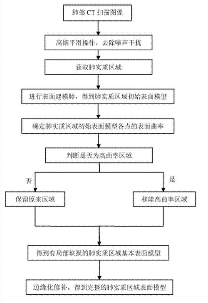
[0025] Such as figure 1 As shown, the implementation process of the modeling method of the present invention is:

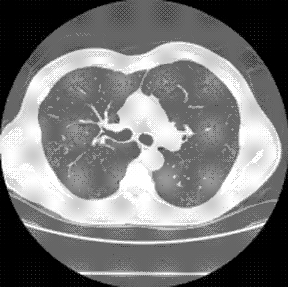
[0026] The first step is to smooth the original image of the lung CT scan image: input a set of DICOM format chest CT slice images containing complete lung organs, a set of 564 original images (the number can be increased or decreased), size 512×512 pixels ( image 3 Is one of them). The Gaussian filter is used to smooth the group of original images to eliminate image noise interference in each image. Since medical images usually contain a lot of noise, which will affect the segmentation results, it is necessary to denoise the image.

[0027] The second step is to obtain the lung parenchymal area: obtain ...


PUM


Abstract

The invention relates to a lung parenchyma area surface model establishment method based on a computed tomography (CT) image. The method includes the steps of conducting gaussian smoothing operation on each original image of a lung CT scanned image by using a computer so as to eliminate image noise interference, obtaining a lung parenchyma area by using a three-dimensional FastMarching algorithm, using a MarchingCubes algorithm, confirming specific morphological characteristics of lung parenchyma by using calculation of surface curvature, removing a confirmed uninterested area, and finally obtaining a complete surface model of a lung interested area with a smooth surface with marginalized repair. The invention provides a computer aided imaging diagnosis method which can eliminate diagnosis difference caused by different subjective factors such as subjective experience and observational ability of doctors, provide referential identified diagnosis results with high accuracy rate, thereby enable diagnostic imaging to be more objective and improve efficiency and accuracy of diagnosis.

Description

Technical field [0001] The invention relates to a method for processing medical images, in particular to a method for establishing a surface model of lung parenchymal region based on CT images. Background technique [0002] Lung cancer is a common primary lung tumor, and its morbidity and mortality are increasing year by year. It has become the malignant tumor with the highest morbidity and mortality in the world. According to the survey results of the Ministry of Health in 2006, my country’s lung cancer mortality rate has increased significantly, and it has become the number one cause of cancer deaths, an increase of 75.77% compared to the 1990s. Excluding the effects of changes in age structure, it has also increased by 33.25%. The clinical manifestations of lung cancer are diverse, and most patients have no obvious symptoms and signs in the early stage, which are difficult to find, and they are often in the late stage. If lung cancer can be diagnosed and treated at an early s...

Claims


Application Information

Patent Timeline
Patent Type & Authority Applications(China)
IPC IPC(8): G06T17/30G06T5/00
Inventor 顾力栩董斌王茜王兵
Owner HEBEI UNIVERSITY
Why Eureka
Try Eureka