Cone-beam X-ray luminescence tomography system of combination imaging and method thereof

An X-ray and cone-beam technology, applied in the electronic field, can solve the problems of long time for collecting fluorescence data, increasing the dose of X-ray irradiation, and X-rays cannot be used to fully excite the medicines of the object to be imaged, so as to overcome the X-ray irradiation time Longer, reduced exposure time and exposure dose, beneficial to popularization and application

Inactive Publication Date: 2013-05-22
XIDIAN UNIV
View PDF11 Cites 25 Cited by
  • Summary
  • Abstract
  • Description
  • Claims
  • Application Information

AI Technical Summary

Problems solved by technology

The disadvantage of this method is that the X-rays emitted by the X-ray source cannot be used to fully excite the medicines

Method used


Image

  • Cone-beam X-ray luminescence tomography system of combination imaging and method thereof
  • Cone-beam X-ray luminescence tomography system of combination imaging and method thereof

Examples


Example Embodiment

[0038] The present invention will be described in detail below with reference to specific embodiments.

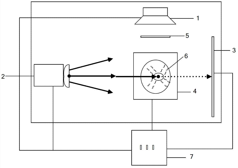
[0039] The present invention is based on a combined imaging cone beam X-ray luminescence tomography imaging system, comprising a CCD camera 1 coupled with a lens, an X-ray source 2, an X-ray detector 3, an electronically controlled rotating table 4, a narrow-band filter 5, and an image to be imaged. Object 6, Computer 7.

[0040] The X-ray source 2 emits X-rays to irradiate the object 6 to be imaged. The medicine in the object 6 to be imaged is excited by the X-rays and emits a fluorescent signal. The fluorescent signal penetrates the object to be imaged 6 , and other signals except the fluorescent signal are filtered out through the narrow-band filter 5 , and the CCD camera 1 receives the transmitted fluorescent signal to generate transmissive fluorescent data. The CCD camera 1 transmits the generated fluorescence projection data to the computer 7 .

[0041] The X-ray s...


PUM


Abstract

The invention discloses a cone-beam X-ray luminescence tomography system of combination imaging and a method thereof. The system comprises a charge-coupled device (CCD) camera, an X-ray source, an X-ray detector, an electronic control rotating table and a narrow-band filter, wherein the CCD camera, the X-ray source, the X-ray detector, the electronic control rotating table and the narrow-band filter are coupled with a lens. The CCD camera and the narrow-band filter are fixed in the same direction, and then a to-be-imaged object, the CCD camera and the narrow-band filter are located on the same straight line; the X-ray source and the X-ray detector are placed in the direction perpendicular to the CCD camera, and then the X-ray source, the to-be-imaged object and the X-ray detector are located on the same straight line; and the CCD camera, the electronic control rotating table, the X-ray source and the X-ray detector are controlled to operate by using a computer. According to the cone-beam X-ray luminescence tomography system of the combination imaging and the method thereof, use ratios of X-rays are improved, and medicine of the to-be-imaged object can be fully excited; X-ray irradiation time is shortened, X-ray irradiation doses are reduced, and popularization and application of X-ray excitation imaging are benefited; information fusion of two types of imaging modals is achieved, and more biological characteristics of the to-be-imaged object are acquired.

Description

technical field [0001] The invention belongs to the field of electronic technology, and particularly relates to a combined imaging cone beam X-ray emission tomography imaging system. The invention utilizes cone beam X-rays to excite the medicines in the object to be imaged, not only can the medicines of the object to be imaged be fully excited, but also the three-dimensional anatomical structure information of the object to be imaged can be obtained by using X-ray imaging, so as to obtain accurate information about the anatomical structure of the object to be imaged. The three-dimensional spatial distribution imaging of drugs realizes multimodal imaging in the process of fluorescence tomography of the object to be imaged. Background technique [0002] X-ray computed tomography (CT) technology is widely used to obtain images of anatomical structures with high spatial resolution, but its role in the field of molecular imaging is very limited. Small amounts of several contrast...

Claims


Application Information

Patent Timeline
IPC IPC(8): A61B6/03
Inventor 朱守平陈冬梅陈多芳屈晓超黄力宇梁继民田捷
Owner XIDIAN UNIV
Why Eureka
Try Eureka