Full-automatic image segmentation method

An image segmentation, fully automatic technology, applied in the field of image processing, can solve problems such as long calculation time, no segmentation method, and difference in segmentation results.

Inactive Publication Date: 2013-07-03
ARMY MEDICAL UNIV
View PDF4 Cites 20 Cited by
  • Summary
  • Abstract
  • Description
  • Claims
  • Application Information

AI Technical Summary

Problems solved by technology

By segmenting cartilage on MRI images and then calculating its thickness, volume and other parameters, the shape of articular cartilage can be quantitatively evaluated. However, the current clinical segmentation of articular cartilage is mainly done manually by radiologists or semi-automatically with the help of image processing software. Not only does it take a long time, but it is inevitable that there will be differences in the segmentation results of different people and at different times
[0003] Due to the complexity of imaging methods, anatomical structures, etc., the segmentation of articular cartilage is always a very challenging task, and there is no general segmentation method at present.
Many researchers have adopted different segmentation strategies for different MRI scan sequences and anatomical planes, and proposed segmentation methods based on theories such as pixel features, edge detection, graph theory, and atlas matching. It is poor, sensitive to noise, and takes a long time to calculate, but it can distinguish different cartilages; the edge-based method cannot distinguish between cartilages that are in contact with each other and muscle tissues with similar gray levels to cartilages, but the edge effect of segmenting cartilages is good. In view of this, It is necessary and meaningful to design a fast, accurate and robust automatic cartilage image segmentation method

Method used


Image

  • Full-automatic image segmentation method
  • Full-automatic image segmentation method
  • Full-automatic image segmentation method

Examples


Embodiment Construction

[0052] The present invention will be described in detail below in conjunction with the accompanying drawings.

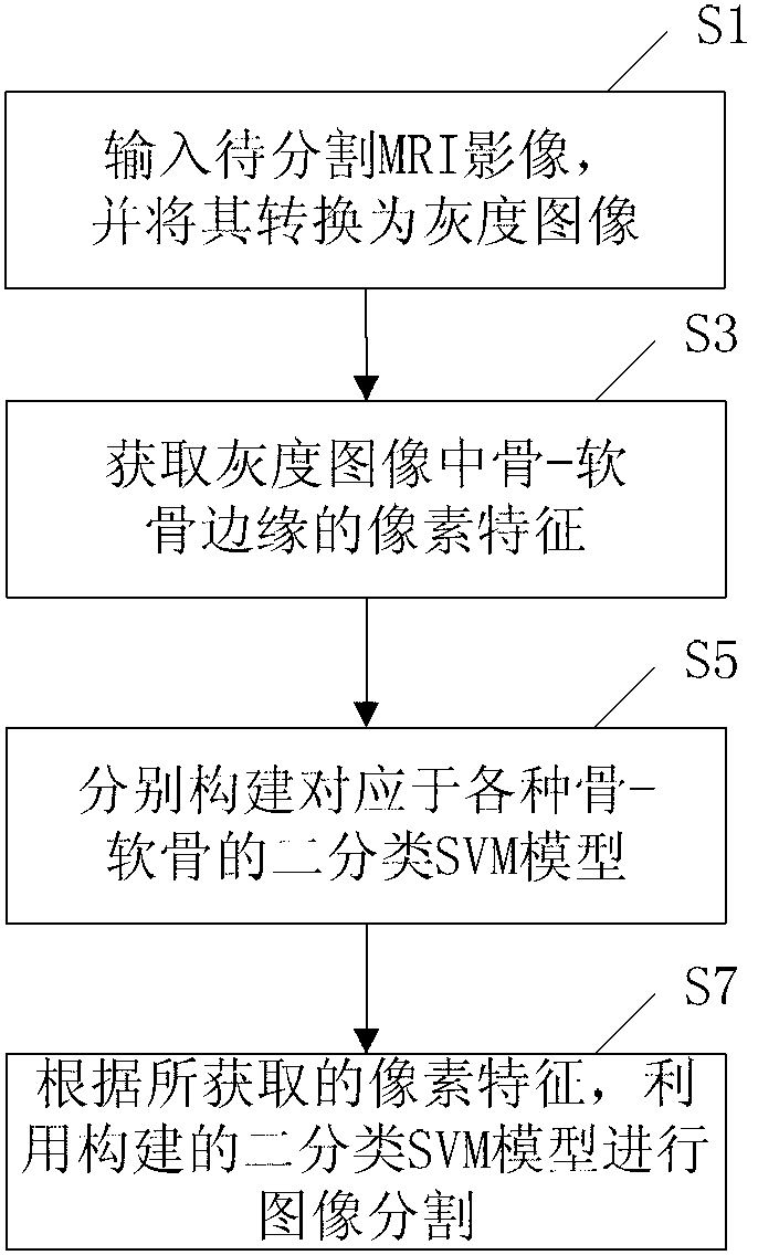
[0053] see figure 1 , is a flowchart of an embodiment of a fully automatic image segmentation method of the present invention. During specific implementation, the fully automatic image segmentation method of this embodiment specifically includes steps:

[0054] S1, input the MRI image to be segmented and convert it into a grayscale image. In one embodiment, the original DICOM image is converted to an 8-bit grayscale image.

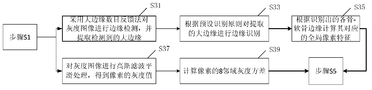
[0055] S3. Obtain pixel features of each bone-cartilage edge in the grayscale image converted in step S1, where the pixel features include global pixel features and local pixel features. In this embodiment, the global pixel feature includes the edge distance and direction of the pixel, and the local pixel feature includes gray value and neighborhood variance. see figure 2 , the step S3 specifically includes:

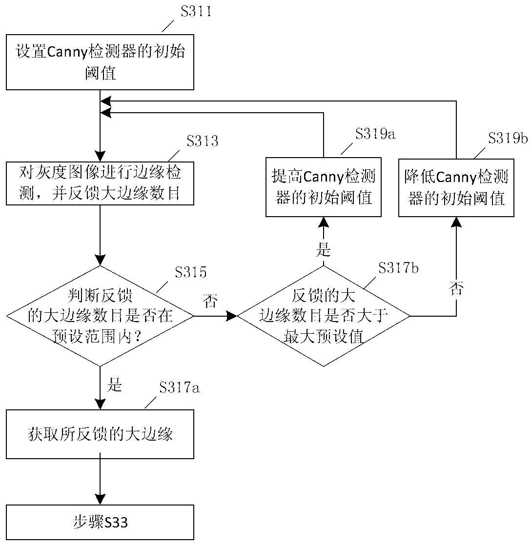
[0056] S31. Using an iterative...


PUM


Abstract

The invention discloses a full-automatic image segmentation method. The method comprises the following steps of: inputting a to-be-segmented MRI (Magnetic Resonance Imaging) image, and converting the image into a gray level image; obtaining pixel feature of each bone-gristle edge in the gray level image, wherein the pixel feature comprises global pixel feature and local feature; respectively forming a binary-class SVM (Support Vector Machine) model corresponding to various bone-gristles; and respectively performing image segmentation for the various bone-gristles in the gray level image through the binary-class SVM model according to the pixel feature. In the method provided by the invention, by obtaining the pixel feature of the bone-gristle edge in the gray level image, and forming corresponding binary-class SVM model for different bone-gristles, the thighbone gristle, the tibia gristle and the patella gristle are independently segmented through the binary-class SVM model according to the obtained pixel feature, pre-segmentation for the bone is not needed, less pixel features are used and the segmentation speed is fast.

Description

technical field [0001] The invention relates to image processing technology, in particular to a fully automatic image segmentation method. Background technique [0002] With the continuous improvement of people's living standards and the intensification of population aging, osteoarthritis has become an important disease that reduces the work and living ability of middle-aged and elderly people. Osteoarthritis is often accompanied by degeneration, destruction and morphological changes of articular cartilage. As a non-invasive and non-invasive examination method, MRI imaging technology has good spatial resolution and tissue contrast, and is an important means to evaluate the morphology and function of cartilage clinically. By segmenting cartilage on MRI images and then calculating its thickness, volume and other parameters, the shape of articular cartilage can be quantitatively evaluated. However, the current clinical segmentation of articular cartilage is mainly done manually...

Claims


Application Information

Patent Timeline
Patent Type & Authority Applications(China)
IPC IPC(8): G06T7/00
Inventor 邱明国庞剑飞陈伟
Owner ARMY MEDICAL UNIV
Why Eureka
Try Eureka