Gene probe composition and kit for detecting epithelial ovarian cancer

A technology of gene probe and composition is applied in the field of gene probe composition and kit for detecting epithelial ovarian cancer, and can solve the problems of low incidence and the like

Active Publication Date: 2013-09-04
INST OF HEMATOLOGY & BLOOD DISEASES HOSPITAL CHINESE ACADEMY OF MEDICAL SCI & PEKING UNION MEDICAL COLLEGE +1
View PDF1 Cites 6 Cited by
  • Summary
  • Abstract
  • Description
  • Claims
  • Application Information

AI Technical Summary

Problems solved by technology

Chk2 gene mutation has certain significance in tumor pathogenesis, but its incidence is low

Method used


Image

  • Gene probe composition and kit for detecting epithelial ovarian cancer
  • Gene probe composition and kit for detecting epithelial ovarian cancer
  • Gene probe composition and kit for detecting epithelial ovarian cancer

Examples


Embodiment 1

[0090] Embodiment 1: The preparation method of BRCA1 gene probe comprises the following steps:

[0091] 1. Clone screening: The BRCA1 gene is located in the 21.2-21.31 segment of the long arm of human chromosome 17 (17q21.2-q21.31), and all clones containing the BRCA1 gene were searched in the UCSC genome browser, NCBI Clone Registry, and Ensembl Genome Browser databases, and screened The optimal clone containing the gene was obtained, numbered RP11-831F13 (as shown in Table 1).

[0092] 2. Cloning culture and identification: purchase clone RP11-831F13 (Invitrogen, USA), take 8 microliters of clone bacteria liquid and add it to 5 milliliters of LB liquid medium containing chloramphenicol resistance, shake and activate at 37°C for 12 hours ; Then add all the bacterial liquid to 450 ml of LB liquid medium containing chloramphenicol resistance, and collect the bacterial liquid after shaking and culturing at 37° C. for 12 hours. Use upstream primer: TAGGGCTGGAAGCACAGAGT (SEQ ID N...

Embodiment 2

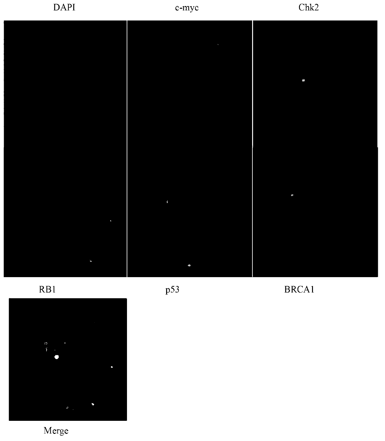
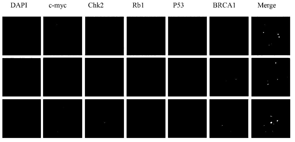
[0119] Embodiment 2: the preparation method of c-myc, Rb1, Chk2, p53, BRCA1 gene probe composition, comprises the following steps

[0120] 1. Clone screening:

[0121] By searching UCSC genome browser, NCBI Clone Registry, Ensembl Genome Browser and other databases contain all clones of c-myc, Rb1, Chk2, p53, BRCA1 genes respectively. The optimal clones containing the gene were screened out, and the numbers were: RP11-440N18, RP11-305D15, RP11-73I4, RP11-1081A10, RP11-831F13. (As shown in table 2)

[0122] 2. Cloning culture and identification: Purchase the clone according to the clone number (Invitrogen, USA), take 8 microliters of the clone bacteria solution and add it to 5 milliliters of chloramphenicol-resistant LB liquid medium, shake and activate at 37°C for 12 hours ; Then add all the bacterial liquid to 450 ml of LB liquid medium containing chloramphenicol resistance, and collect the bacterial liquid after shaking and culturing at 37° C. for 12 hours. The bacteria l...

Embodiment 3

[0160] Example 3: A fluorescent in situ hybridization detection kit for epithelial ovarian cancer (50 servings), the composition is as follows:

[0161] 1) Fluorescence-labeled probe set hybridization mixture 100 microliters; 1 tube

[0162] 2) DAPI counterstain solution 500 microliters; 1 tube

[0163] 3) 20ml of 20XSSPE washing solution; 1 bottle

[0164] 4) 1 instruction manual.

[0165] Wherein the concentration of the c-myc gene probe in the hybridization mixture of the fluorescently labeled probe group is: 4ng / μl, the concentration of the Rb1 gene probe is: 4ng / μl, the concentration of the Chk2 gene probe is: 4ng / μl, and the concentration of the p53 gene probe 4ng / μl, BRCA1 gene probe concentration: 4ng / μl.


PUM

PropertyMeasurementUnit
molecular weightaaaaaaaaaa

Abstract

The invention relates to a gene probe composition and a kit for detecting epithelial ovarian cancer. The gene probe composition comprises a c-myc gene probe, an Rb1 gene probe, a Chk2 gene probe, a p53 gene probe and a BRCA1 gene probe. Another objective of the invention is to provide a kit for fluorescence in situ hybridization (FISH) detection of the epithelial ovarian cancer, and the kit comprises the gene probe composition as described above. The kit can detect 5 kinds of genes in a same sample even a single tumor cell of the epithelial ovarian cancer at the same time, and thereby substantially improving detection capability and efficiency. The invention further provides an optimal sample selection strategy and a slide preparation method for an ovarian cancer FISH research.

Description

technical field [0001] The invention relates to a gene probe composition and a kit, in particular to a gene probe composition and a kit for detecting epithelial ovarian cancer. Background technique [0002] Ovarian cancer is one of the most common tumors in female reproductive organs, ranking third in incidence after cervical cancer and uterine body cancer. However, the number of deaths due to ovarian cancer ranks first in all types of gynecological tumors, posing a serious threat to women's lives. The etiology of ovarian cancer is still unclear. Ovarian cancer has atypical clinical symptoms due to hidden anatomical sites. About 70% of ovarian cancer patients are already in the advanced stage (III, IV stage) when ovarian cancer is discovered. In recent years, combined with tumor reduction surgery With the development of chemotherapy, the survival rate of patients has improved, but the overall situation is not optimistic. About 80% to 85% of ovarian cancer patients will rel...

Claims


Application Information

Patent Timeline
Patent Type & Authority Applications(China)
IPC IPC(8): C12Q1/68C12N15/11C12N15/10
Inventor 程涛黄宇婷胡林萍郝权
Owner INST OF HEMATOLOGY & BLOOD DISEASES HOSPITAL CHINESE ACADEMY OF MEDICAL SCI & PEKING UNION MEDICAL COLLEGE
Why Eureka
Try Eureka