Active contour model based method for segmenting mammary gland DCE-MRI focus

A technology of DCE-MRI and active contour model, applied in image analysis, image data processing, instruments, etc., can solve the problems of fuzzy lesion boundaries, inability to cover the overall situation of lesions, and lack of statistical characteristics

Active Publication Date: 2013-10-02
DALIAN UNIV OF TECH
View PDF4 Cites 25 Cited by
  • Summary
  • Abstract
  • Description
  • Claims
  • Application Information

AI Technical Summary

Problems solved by technology

Shannon C. Agner et al. proposed a hybrid active contour model method to segment two-dimensional lesions. This algorithm combines the manifold method to convert the image space to the spectral space to improve the blurred edge of the lesion in the original space. Due to the computational spectral space The tensor gradient in it takes a long time, which will limit its real-time performance for processing complex images
In addition, in most methods based on the active contour model, in order to ensure the stability of the numerical calculation of the algorithm, the initialization of the signed distance function needs to be repeated during the evolution of the curve or surface, which takes a long time, which greatly limits the real-time application of the algorithm.
[0004] Summarizing the current research on the segmentation of lesions in dynamic contrast-enhanced breast magnetic resonance imaging, it can be found that there are several difficulties: (1) The lesions are adjacent to normal tissues, and the boundaries of the lesions are blurred. If they cannot be effectively distinguished, the segmentation will easily cause boundary leakage; 2) The distribution of gray levels inside the lesion is diverse, which is effective information for lesion staging and diagnosis. It does not have consistent statistical characteristics, and conventional modeling often cannot cover the overall situation of the lesion; (4) In order to obtain comprehensive and three-dimensional spatial information of lesions, three-dimensional angle segmentation is very necessary. However, DCE-MRI images have a large amount of data, and unreasonable segmentation process will result in long time and low efficiency of segmentation calculation.

Method used


Image

  • Active contour model based method for segmenting mammary gland DCE-MRI focus
  • Active contour model based method for segmenting mammary gland DCE-MRI focus
  • Active contour model based method for segmenting mammary gland DCE-MRI focus

Examples


Embodiment Construction

[0051] The implementation method of the present invention will be described in further detail below in combination with examples and accompanying drawings.

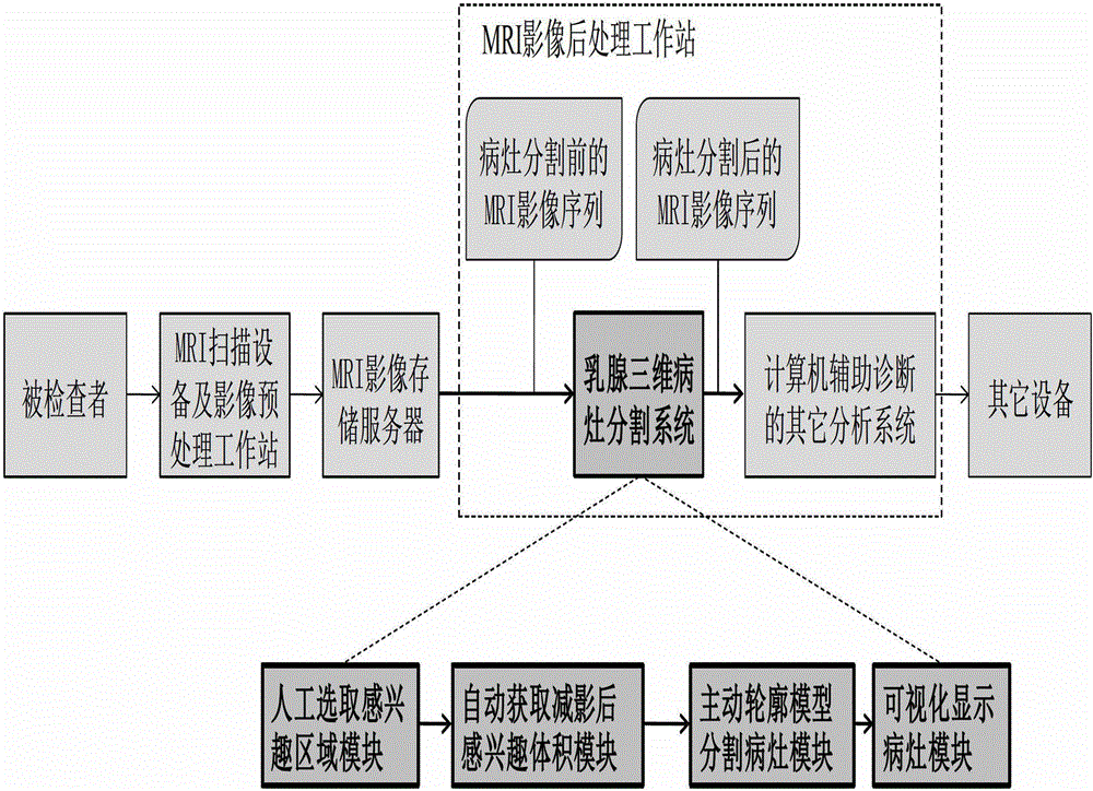
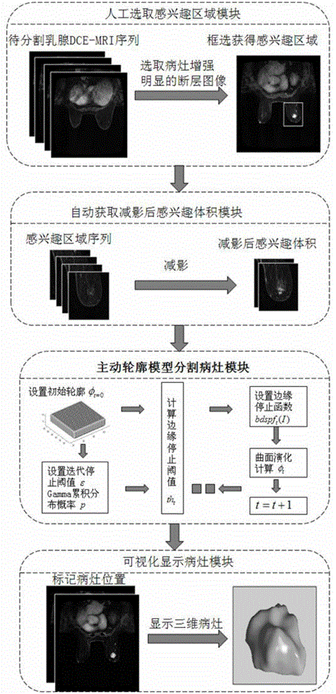
[0052] In order to implement the breast DCE-MRI three-dimensional lesion segmentation method proposed by the present invention, it needs to follow the following steps: figure 1 In the process shown, the breast MRI image sequence data of the examinee is obtained from the MRI scanning equipment and screened by the image preprocessing workstation, and the filtered data is saved in the MRI image storage server used to save all the examinee image sequences, and then The MRI image post-processing workstation obtains the image sequence to be processed from the MRI image storage server for analysis and calculation. The construction of the three-dimensional breast lesion segmentation system in the MRI image post-processing workstation includes the module of manually selecting the region of interest, the module of automatically obt...


PUM


Abstract

An active contour model based method for segmenting mammary gland DCE-MRI focus belongs to the field of medical image segmentation and comprises the following steps: obtaining mammary gland DCE-MRI image sequence data by MRI scanning equipment; manually selecting a region of interest; automatically obtaining subtracted size of interest, active contour segmenting focus and visually display focus. According to the invention, based on the features that statistical distributions of mammary gland DCE-MRI image backgrounds are consistent and internal distributions in the focus are different, an edge stopping function of the active contour model is designed, thereby realizing reliable segmentation of the focus and effectively avoiding edge outleakage phenomenon; during the model evolutionary process, re-initialization of a signed distance function is not required, so that the real-time performance of the system is higher. The method has a lower requirement on manual operation in implementation, is high in intelligent degree, low in data storage space requirement, and quick in processing speed, and can effectively obtain comprehensive and steric space information of the focus through three-dimensional angle segmentation, which facilitates the multi-angle observation and analysis of the focus by a doctor.

Description

technical field [0001] The invention relates to the technical field of medical image segmentation, in particular to an active contour model method for rapidly and reliably segmenting three-dimensional lesions in breast dynamic contrast-enhanced magnetic resonance image sequences based on level set theory. Background technique [0002] In recent years, breast DCE-MRI scanning has become one of the most potential imaging techniques for detecting breast diseases, and plays an important role in the clinical diagnosis and medical research of early breast cancer. At the same time, the DCE-MRI-based computer aided diagnosis (computer aided diagnosis, CAD) system has been gradually applied to in clinical practice. Accurate and reliable lesion segmentation not only marks the detection of lesions, but also restricts the accuracy of subsequent morphological, texture, and hemodynamic feature extraction, thereby affecting the correctness of diagnostic conclusions. Therefore, lesion segm...

Claims


Application Information

Patent Timeline
Patent Type & Authority Applications(China)
IPC IPC(8): G06T7/00
Inventor 刘惠柳怡萍
Owner DALIAN UNIV OF TECH
Why Eureka
Try Eureka