Method for preparing to-be-repaired skull flap by 3D printing

A 3D printing and bone flap technology, applied in 3D modeling, bone implants, medical science, etc., can solve the problems of insufficient strength, poor heat preservation, high cost, etc., and achieve beautiful appearance, high safety and reliability , the effect of low price

Active Publication Date: 2013-11-20
TONGJI HOSPITAL ATTACHED TO TONGJI MEDICAL COLLEGE HUAZHONG SCI TECH
View PDF12 Cites 37 Cited by
  • Summary
  • Abstract
  • Description
  • Claims
  • Application Information

AI Technical Summary

Problems solved by technology

[0007] The purpose of the present invention is to overcome the defects of existing cranial repair methods, such as the difference between the repaired skull flap made of titanium alloy mesh and the

Method used


Image

  • Method for preparing to-be-repaired skull flap by 3D printing

Examples


Embodiment Construction

[0030] The present invention will be further described in detail below in conjunction with the accompanying drawings and specific embodiments.

[0031] A method for preparing a skull flap to be repaired by 3D printing, the steps are as follows:

[0032] 1) Use a photographic 3D scanner to perform three-dimensional scanning of the original bone flap to be repaired at the skull defect, and obtain the three-dimensional data of the physical surface points of the original bone flap;

[0033] The original bone flap needs preliminary treatment before scanning. The original bone flap removed during the operation is soaked in 10% formalin solution for 4 hours, rinsed with clean water for 3 times, and the connective tissue attached to the surface of the skull flap is scraped off. The tissue is ready for the next scan. The cleaned original skull bone flap can also be placed in a drying oven, baked at 60°C for 6 hours, and kept at room temperature for a long time for future scanning.

...


PUM

PropertyMeasurementUnit
Apertureaaaaaaaaaa

Abstract

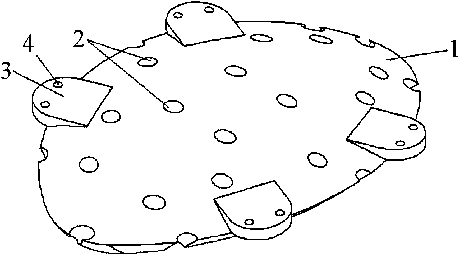
The invention discloses a method for preparing a to-be-repaired skull flap by 3D printing. The method comprises the following steps: three-dimensionally scanning an original flap at a to-be-repaired skull defect part to obtain the three-dimensional data of surface points of an original flap entity; generating a skull flap three-dimensional model matched with the original flap; forming 8-16 drainage holes in the model and circumferentially and uniformly arranging a plurality of tabs protruding from the skull flap edge along the skull flap three-dimensional model; taking medical photosensitive resin as a raw material, and printing the raw material by a 3D printer to obtain a medical resin flap; finally, fixing the resin flap and the to-be-repaired skull by a tapping screw. Through adoption of the method, the medical resin flap matched with the height of the skull defect window of a patient can be prepared, secondary molding is not required in operation, and the operation time is shortened. The medical resin flap is light in weight, solid, excellent in impact resistance, low in heat-conducting property, and heat-insulating, and reduces discomfort caused after head operation.

Description

technical field [0001] The invention relates to a method for preparing a skull bone flap for repair, in particular to a method for preparing a skull bone flap to be repaired by using 3D printing. Background technique [0002] Skull defects caused by various reasons are very common in clinical practice, especially in the treatment of severe craniocerebral trauma. The guidelines for the treatment of craniocerebral trauma in various countries recommend standardized decompression of large cranial craniectomy combined with evacuation of intracranial hematoma as the preferred treatment method , because the removal of the skull flap can very effectively reduce the intracranial pressure, so as to reduce the compression on the life center of the brainstem and save the patient's life. Therefore, after a large number of patients with severe craniocerebral trauma are surgically treated, many patients with skull defects will be left clinically. [0003] Although the removal of the skull...

Claims


Application Information

Patent Timeline
IPC IPC(8): A61F2/28A61L27/14G06T17/00
CPCA61F2/28A61F2002/30001
Inventor 唐朝晖唐庭轩余彦
Owner TONGJI HOSPITAL ATTACHED TO TONGJI MEDICAL COLLEGE HUAZHONG SCI TECH
Why Eureka
Try Eureka