Method for extracting stomach CT (Computed Tomography) image suspected to be lymph node based on sparse dynamic integrated selection

A CT image and extraction method technology, applied in the field of medical image processing, can solve problems such as high computational complexity and time-consuming, achieve good classification performance and reduce computational complexity

Active Publication Date: 2013-11-20
XIDIAN UNIV
View PDF3 Cites 6 Cited by
  • Summary
  • Abstract
  • Description
  • Claims
  • Application Information

AI Technical Summary

Problems solved by technology

Dynamic ensemble selection is the latest achievement of multi-classifier ensemble. It has received extensive attention because it can achieve better classification performance. However, in the prior art, when dynamic ensemble selection is used to classify adipose tissue, the computational complexity is high. takes a lot of time

Method used


Image

  • Method for extracting stomach CT (Computed Tomography) image suspected to be lymph node based on sparse dynamic integrated selection
  • Method for extracting stomach CT (Computed Tomography) image suspected to be lymph node based on sparse dynamic integrated selection
  • Method for extracting stomach CT (Computed Tomography) image suspected to be lymph node based on sparse dynamic integrated selection

Examples


Embodiment 1

[0046] The present invention is a sparse dynamic integration selection method for extracting suspected lymph nodes from gastric CT images. Currently, CT images are commonly used imaging methods. To realize the extraction of suspected lymph node areas in fat tissue areas of CT images, the fat tissue areas must be extracted first. . Lymph nodes are mainly distributed in the adipose tissue around the stomach wall, so accurate segmentation of adipose tissue is of primary importance.

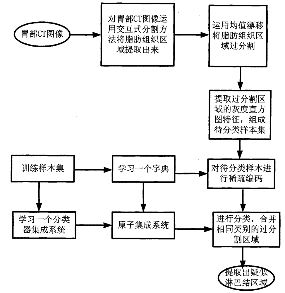
[0047] In the present invention, the process of extracting suspected lymph node regions from images of gastric adipose tissue is as follows: figure 1 shown. The specific description is as follows:

[0048] Step 1, extract a CT image of the stomach, see Figure 4 (a). The stomach CT images used in this example are from Beijing Cancer Hospital. Each CT image consists of 512x512 pixels. All image processing experiments are implemented on the MATLAB2009a experimental platform.

[0049] Step 2, artif...

Embodiment 2

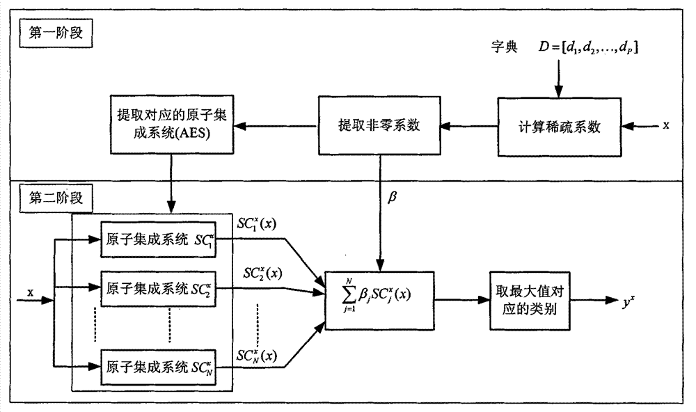
[0066] The method for extracting suspected lymph nodes from gastric CT images selected by sparse dynamic integration is the same as in Example 1, wherein the process of using the sparse dynamic integration selection method described in step 5 to classify the extracted adipose tissue region sample set to be classified includes a training phase and a test stage:

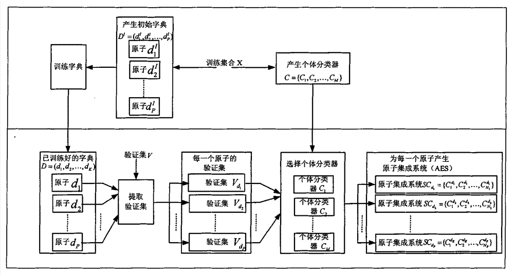
[0067] The flow chart of the training phase is as follows figure 2 As shown, the specific process is:

[0068] 5.1 Select training samples from some marked images of adipose tissue that have been successfully classified, and the training samples are denoted as X={(x i ,y i )|x i ∈ R F ,y i ∈ {1, 2, ..., L}, i = 1, 2, ..., r}, where x i is the grayscale histogram feature extracted from the labeled image of adipose tissue, y i is the label of the sample, F is the dimension of the proposed gray histogram feature, L is the number of categories to be segmented, and r is the number of samples to be extracted; the num...

Embodiment 3

[0091] The method for extracting suspected lymph nodes from gastric CT images selected by sparse dynamic integration is the same as in Embodiment 1-2. In this example, the effectiveness of the present invention is verified by simulation experiments using gastric CT images from Beijing Cancer Hospital. Each image consists of 512x512 pixels, and three images are selected for experiments. All experiments are implemented on the MATLAB2009a experimental platform. Figure 4-Figure 6 Adipose tissue extraction results for three images. exist Figure 4-Figure 6 Among them, for each picture, (a) is the original image, (b) is the result after inputting the interactive information, (c) is the accurate fat tissue area extraction result, (e) is the fat tissue obtained by the method used in the present invention The results of tissue region extraction, (d) is the partial enlarged image corresponding to (c), and (f) is the partial enlarged image corresponding to (e). Figure 4-Figure 6 The ...


PUM


Abstract

The invention discloses a method for extracting a stomach CT (Computed Tomography) image suspected to be a lymph node based on sparse dynamic integrated selection and belongs to the field of medical image processing. According to the method, the problems that effective background information is difficult to determine and is high in complexity when a suspected lymph node area is extracted are mainly solved. The realization process of the method comprises the following steps of extracting an adipose tissue area from the stomach CT image by using an interactive segmentation method; over segmenting the extracted adipose tissue area by using mean shift; extracting the characteristics of a grey level histogram in the over-segmentation area to form a sample set to be classified; utilizing a training sample learning dictionary and a classifier integration system; generating an atom integration system for each atom; sparsely encoding a sample to be classified and outputting a final classifying result according to the atom integration system; combining the over-segmentation areas of the same type to obtain the stomach CT image suspected to be the lymph node area. According to the method disclosed by the invention, adipose tissues can be automatically extracted under the condition that no background is marked and the time complexity of extraction of the suspected lymph node area is effectively reduced.

Description

technical field [0001] The invention belongs to the technical field of medical image processing, in particular to segmentation and classification of medical CT images, in particular to a method for extracting suspected lymph nodes from gastric CT images with sparse dynamic integration selection. It can be used for segmentation and classification of CT images of gastric adipose tissue, can be used for segmentation and classification of other types of CT images, and can also be used for segmentation or classification of other types of images. Background technique [0002] Gastric cancer ranks second in the mortality rate of malignant tumors in the world, and its rapid development and easy metastasis are important reasons for the high mortality rate. Lymph node metastasis is an important independent factor affecting the prognosis of gastric cancer. Before medical and surgical treatment, it is necessary to know the lymph node status as accurately as possible to determine the tre...

Claims


Application Information

Patent Timeline
Patent Type & Authority Applications(China)
IPC IPC(8): G06T7/00A61B6/03
Inventor 刘芳周治国李玲玲郝红侠戚玉涛焦李成李婉尚荣华马文萍马晶晶
Owner XIDIAN UNIV
Why Eureka
Try Eureka