
System and method for ultrasound examination of the breast

An ultrasonic imaging and ultrasonic sensor technology, applied in ultrasonic/sonic/infrasonic Permian technology, organ motion/change detection, ultrasonic/sonic/infrasonic diagnosis, etc., can solve problems such as uneven distance

Inactive Publication Date: 2015-05-20
SONARIUM MEDICAL
View PDF13 Cites 0 Cited by
  • Summary
  • Abstract
  • Description
  • Claims
  • Application Information

AI Technical Summary

Problems solved by technology

Also, because the transducer's annulus has a fixed diameter, as the breast is scanned, the distance between the annulus and the breast is not uniform due to the taper of the breast toward the nipple

Method used


Image

  • System and method for ultrasound examination of the breast
  • System and method for ultrasound examination of the breast
  • System and method for ultrasound examination of the breast

Examples


Embodiment Construction

[0175] For clarity and ease of description, the present invention will be described with respect to breast imaging, it being apparent that the systems and methods of the present invention may be modified to image any desired body part.

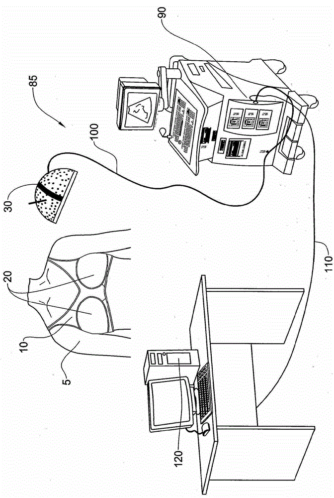
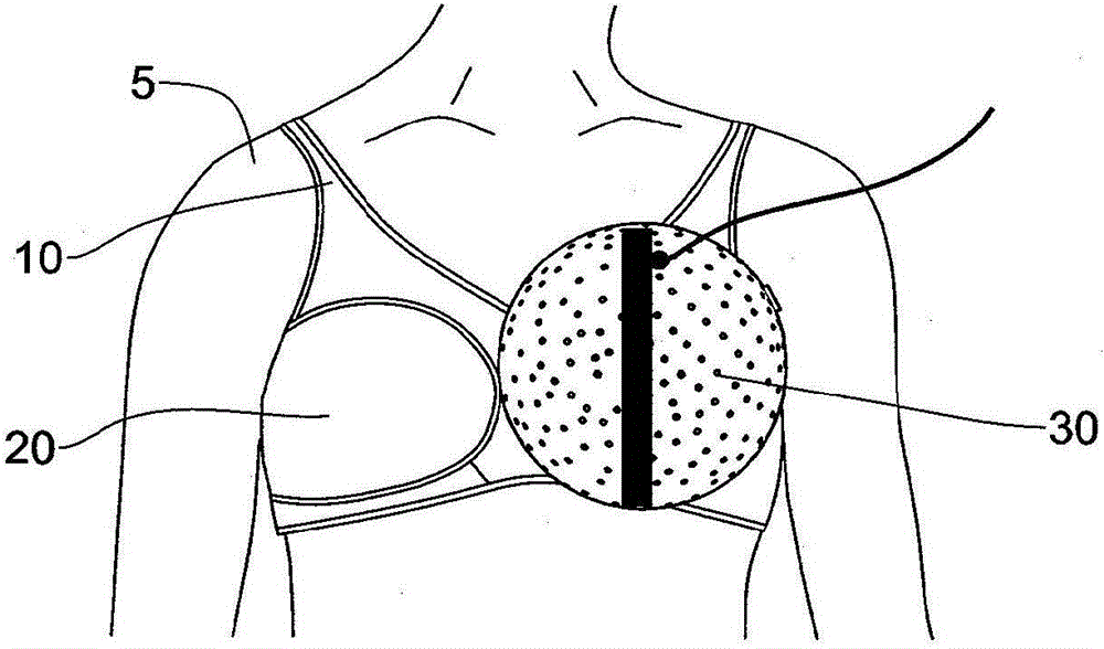
[0176] figure 1 A system 85 for ultrasound imaging of a breast according to one embodiment of the invention is shown. The system 85 includes a dome-shaped scanning device 30, described in detail below, configured to receive the breast of the subject 5 therein. Scanning device 30 is secured to ultrasound system 90 by cable assembly 100 . A control cable 110 connects the ultrasound system 90 to a workstation 120 . Workstation 120 may include a CRT screen 123 for displaying images. User input means such as a keypad 124 allow the user to enter various parameters related to the examination, such as personal details of the subject or parameters of the ultrasound radiation (frequency, intensity, etc.).

[0177] In one embodiment of the present in...


PUM


Abstract

The present invention provides a system and method for limited field of view ultrasound imaging of a 2D section or 3D volume of a body part. The ultrasonic sensors are configured to be spatially or temporally arranged in a restricted field of view arc or on at least a portion of a concave surface such as a hemisphere. The processor calculates a beamforming (BF) function from the detected ultrasound radiation and a point spread function (PSF) from the free amplitude. Fourier transform H from the PSF of the image used to generate a 2D section or 3D volume of a body part BF (k) Calculate the filter g(k).

Description

technical field [0001] The present invention relates to medical devices, and more particularly to devices for medical imaging by ultrasound. Background technique [0002] The following prior art disclosures are considered relevant for understanding the prior art. [0003] 1. Report on "Mammography and beyond: developing technologies for the early detection of breast cancer", [0004] 2. U.S.A. Institute of Medicine and the Governing Board of the National Research Council, National Academy Press, 2101 Constitution Avenue, N.W., Box 285, Washington, DC 20055. [0005] 3. Chandra M. Sehgal et al., Journal of Mammary Gland Biology and Neoplasia, April 2006, Volume 11, Issue 2. [0006] 4. W.E. Svensso, "Breast Ultrasound Update", ULTRASOUND, Vol. 14, No. 1, Feb. 2006, pp. 22-30. [0007] 5. Quantitative cross-sectional imaging of ultrasound parameters by J.F. Greenleaf et al., Ultrasonics Symposium Proc, IEEE Cat.#77CH1264-1SU, pp. 989-995, 1977. [0008] 6. Littrup PJ et al...

Claims


Application Information

Patent Timeline
Patent Type & Authority Patents(China)
IPC IPC(8): A61B8/08A61B8/13
CPCA61B8/0825A61B8/406A61B8/4477A61B8/466A61B8/4461A61B8/461
Inventor M·贝尔曼
Owner SONARIUM MEDICAL
Learn More