Intracavitary ultrasonic lithotripsy device

A technique of intracavity ultrasound and lithotripsy, applied in the direction of internal fixator, internal bone synthesis, application, etc., can solve the problems of increasing patient trauma, pain, visual field interference, and prolonging operation time, so as to improve the operation efficiency and effect, Ease of pain and burden, and ease of crushing stones

Inactive Publication Date: 2013-12-11
HENAN UNIV OF SCI & TECH
View PDF4 Cites 5 Cited by
  • Summary
  • Abstract
  • Description
  • Claims
  • Application Information

AI Technical Summary

Problems solved by technology

However, at present, endoscopic stone removal often has the following disadvantages: 1. Larger single stones need to be crushed and removed in stages, and the visual field is disturbed, unclear, and stone debris remains during the stone removal process, which may cause recurrence of stones after surgery , surrounding tissue damage and other complications
2. For multiple stones, the above disadvantages are more obvious, and the operation time is significantly prolonged due to low stone removal efficiency, unclear visual field interference, and residual stones, and the stone removal effect is not good, which increases the risk of surgery and anesthesia, so it has to be changed. For open surgery, increase the trauma and pain of the patient

Method used


Image

  • Intracavitary ultrasonic lithotripsy device
  • Intracavitary ultrasonic lithotripsy device
  • Intracavitary ultrasonic lithotripsy device

Examples


Embodiment Construction

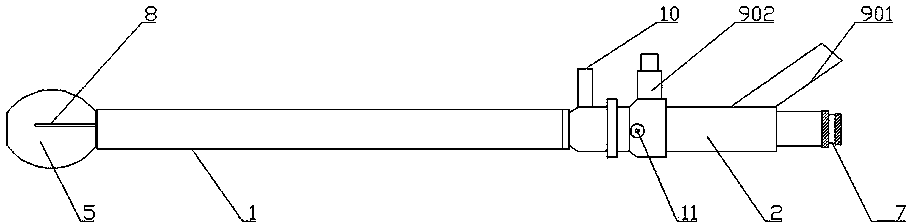
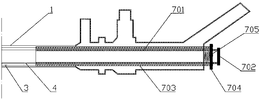
[0027] Such as figure 1 , 2 Shown in , 3: an intracavity ultrasonic lithotripsy device, comprising a straight tubular outer mirror sheath 1 open at both ends and a mirror body 2 connected to one end of the outer mirror sheath 1, the outer mirror sheath 1 is provided with a lithotripsy assembly 8 and As for the endoscopic imaging component, the lithotripsy component 8 is an ultrasonic lithotripsy component, and the lithotripsy component 8 includes an ultrasonic generator and a lithotripsy rod. The outer mirror sheath 1 is provided with a rotating sheath 3 and an endoscope sheath 4, the rotating sheath 3 and the endoscope sheath 4 are straight tubular mirror sheaths with open ends, the rotating sheath 3 can be rotatably sleeved outside the endoscope sheath 4, so The inner wall of the rotating sheath 3 is provided with an annular groove 301, and the outer wall of the endoscope sheath 4 is provided with a tab 401 that snaps into the annular groove 301, and there is no axial rela...


PUM


Abstract

The invention relates to an intracavitary ultrasonic lithotripsy device. The intracavitary ultrasonic lithotripsy device comprises an outer lens sheath and a lens body, wherein a rotating sheath and an inner lens sheath are arranged in the outer lens sheath, and the inner lens sheath is sheathed in the rotating sheath in a rotating manner and no relative displacement in the axial direction is generated between the rotating sheath and the inner lens sheath; a stone pickup assembly is arranged on one end, away from the lens body, of the inner lens sheath; an opening is formed in the side wall of the stone pickup assembly and can be covered by a sealing assembly, an instrument channel tube is arranged in the cavity of the inner lens sheath, a stone breaking assembly is arranged in the instrument channel tube, and a stone breaking end of the stone breaking assembly stretches into the stone pickup assembly. According to the intracavitary ultrasonic lithotripsy device, stones can be limited in a sealed space, stone residues cannot enter a human body when the lithotripsy is carried out, no stone is left after the lithotripsy, the damage to surrounding tissues is little, and the interference caused by the stone chippings with the view of an endoscope is little, so that the operation efficiency and effect can be remarkably improved, and the pain and burden of a patient are obviously relieved.

Description

technical field [0001] The invention relates to the technical field of medical instruments, in particular to an intracavity ultrasonic lithotripsy device. Background technique [0002] Stone disease mostly occurs in the urinary system and biliary system, and is a common and frequently-occurring disease. It is mainly treated by extracorporeal shock wave lithotripsy (ESWL), endoscopic stone extraction and open surgery. Because extracorporeal shock wave lithotripsy is affected by various interference factors, including the patient's physique, stone quantity, location, activity, and whether it adheres to surrounding tissues, etc., the scope of use and the effect of lithotripsy are greatly reduced. Clinical studies have shown that although extracorporeal shock wave lithotripsy has the characteristics of non-invasiveness and repeated strong operations, repeated lithotripsy on one part often leads to adhesion of stones and surrounding tissues, which must be resolved by open surgery...

Claims


Application Information

Patent Timeline
IPC IPC(8): A61B17/22A61B17/94
Inventor 时振国朱小娟
Owner HENAN UNIV OF SCI & TECH
Why Eureka
Try Eureka