Multiple modality cardiac imaging

A diagnostic imaging and blood vessel image technology, which is applied in the field of medical imaging, can solve problems such as the inability to generate attenuation correction maps, and achieve the effects of shortening the total inspection time, reducing data quality, and reducing risks

Inactive Publication Date: 2013-12-18
KONINKLJIJKE PHILIPS NV
View PDF9 Cites 11 Cited by
  • Summary
  • Abstract
  • Description
  • Claims
  • Application Information

AI Technical Summary

Problems solved by technology

Therefore, accurate untruncated attenuation correction maps

Method used


Image

  • Multiple modality cardiac imaging
  • Multiple modality cardiac imaging
  • Multiple modality cardiac imaging

Examples


Embodiment Construction

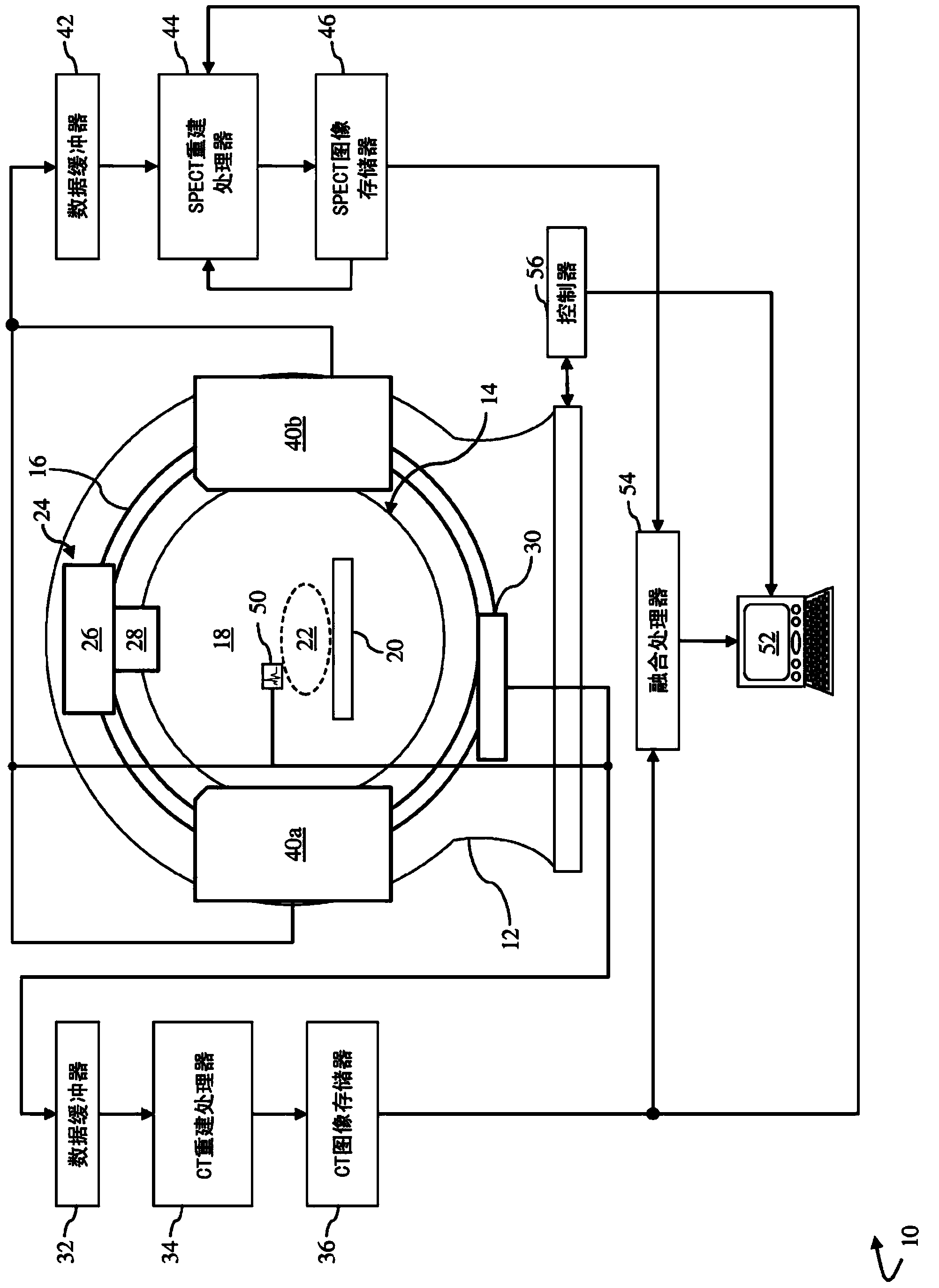
[0027] A multimodal diagnostic imaging system capable of x-ray CT and nuclear imaging acquisitions enables comprehensive assessment of coronary artery disease using a single x-ray CT acquisition. Generate cardiac perfusion SPECT reconstructions, 2D x-ray angiograms and 3D x-ray coronary reconstructions during a single imaging session. The system reduces exam cost and overall exam time by generating more comprehensive exams available to more patients, and by reducing the risk of misregistration and misdiagnosis between imaging exams due to patient motion and anatomical variation , which improves the diagnosis of coronary artery disease. The system is not limited to cardiac imaging studies, and may be applicable to study various vasculature studies throughout a patient's body, for example, to study the nervous system. refer to figure 1 , the diagnostic imaging system 10 performs simultaneous and / or independent x-ray computed tomography (CT) and nuclear imaging such as PET or S...


PUM


Abstract

A multiple modality imaging system 10 for cardiac imaging includes x-ray scanners 24,30 which acquires contrast enhanced CT projection data of coronary arteries with a laterally offset flat panel detector and SPECT imaging scanners 40a,40b, which share the same examination region and gantry as the x-ray scanners, acquires nuclear projection data of the coronary arteries. A CT reconstruction processor 34 generates a 3D coronary artery image representation, at least one planar coronary artery angiogram, and a 3D attenuation correction map from the acquired CT projection data. A SPECT reconstruction processor 44 corrects the acquired nuclear projection data based on the generated attenuation correction map and generates a SPECT image representation of the coronary arteries from the corrected nuclear projection data. A fusion processor 54 combines the nuclear image representation, the 3D vessel image representation, and the at least one planar vessel angiogram into a composite image.

Description

technical field [0001] This application relates to the field of medical imaging. It is particularly useful for coronary artery imaging and perfusion studies using multimodal imaging systems that perform both nuclear and x-ray / CT imaging. Background technique [0002] In diagnostic nuclear imaging, radionuclides are passed through a patient's bloodstream, with this passage being studied for radionuclide distribution to image the circulatory system or to image specific organs where injected radiopharmaceuticals accumulate. Advantageously, the radiopharmaceutical can be designed to concentrate in selected tissues to provide preferential imaging of these selected tissues. [0003] In single photon emission computed tomography (SPECT), one or more radiation detectors, collectively known as gamma cameras, are used to detect radiopharmaceuticals through the emission of radiation from radioactive decay events. Typically, each gamma camera includes an array of radiation detectors a...

Claims


Application Information

Patent Timeline
IPC IPC(8): A61B6/03
CPCA61B6/032A61B6/037A61B6/4417A61B6/4429A61B6/503A61B6/506A61B6/5205A61B6/5235A61B6/5258A61B6/5288G06T11/006G06T2211/404G06T2211/412G06T2211/432A61B6/4258A61B6/481A61B6/035A61B6/463A61B6/466A61B6/504
Inventor E·S·汉西斯
Owner KONINKLJIJKE PHILIPS NV
Why Eureka
Try Eureka