Method and device for abdomen soft tissue nuclear magnetism image segmentation

A nuclear magnetic image and soft tissue technology, which is applied to the field of organ tissue segmentation algorithms for abdominal nuclear magnetic images, can solve problems such as inability to obtain results, difficulty in separate segmentation of medical image grayscale changes, and achieve the effect of simple algorithm implementation.

Active Publication Date: 2013-12-25
SHENZHEN INST OF ADVANCED TECH CHINESE ACAD OF SCI
View PDF3 Cites 5 Cited by
  • Summary
  • Abstract
  • Description
  • Claims
  • Application Information

AI Technical Summary

Problems solved by technology

[0009] The existing image algorithm only relies on the gray information of the image to segment, and cannot obtain ideal results, and the complexity of medical images and the characteristics of little gray change between different tissues make it very difficult to se

Method used


Image

  • Method and device for abdomen soft tissue nuclear magnetism image segmentation
  • Method and device for abdomen soft tissue nuclear magnetism image segmentation
  • Method and device for abdomen soft tissue nuclear magnetism image segmentation

Examples


Embodiment Construction

[0045] In order to make the object, technical solution and advantages of the present invention more clear, the present invention will be further described in detail below in conjunction with the examples. It should be understood that what is described here is only a part of the embodiments of the present invention, rather than all the embodiments. Based on the embodiments of the present invention, all other embodiments obtained by persons of ordinary skill in the art without making creative efforts belong to the protection scope of the present invention.

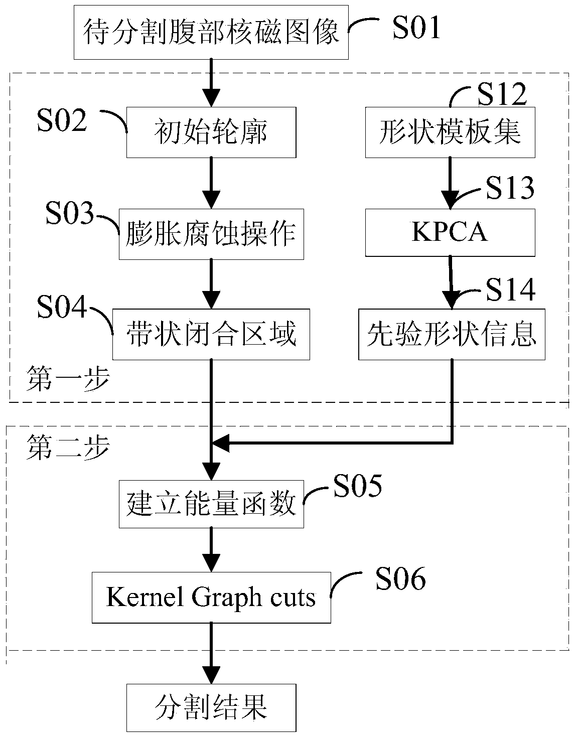
[0046] One of the purposes of the embodiments of the present invention is to provide a method for segmenting an abdominal soft tissue nuclear magnetic image, which is used to improve the robustness of the nuclear magnetic image segmentation algorithm and make the segmentation result more accurate. In order to achieve the above purpose, the method for the segmentation of abdominal soft tissue MRI images, such as figure 1 sho...


PUM


Abstract

The invention discloses a method and device for abdomen soft tissue nuclear magnetism image segmentation. The method comprises the steps that an original outline is initialed near an objective outline; a morphological operator is used for carrying out expansion and corrosion operations on the original outline, and a band-shaped closed area is formed in and outside the objective outline to be segmented; KPCA training is conducted on a collected shape template and prior shape information is obtained through a statistics model; the prior shape information is combined with data items of an energy function of a nuclear magnetism image segmentation model, and an energy function is constructed; a kernel Graph cuts algorithm is used for carry out segmentation on the band-shaped closed area and an objective outline is obtained. The method and device for abdomen soft tissue nuclear magnetism image segmentation can achieve semi-automatic segmentation, the device is simple, the robustness of the nuclear magnetism image segmentation algorithm is effectively improved so as to enable the segmentation result to be more accurate, and the method and device for abdomen soft tissue nuclear magnetism image segmentation can be applied to most nuclear magnetism image segmentation.

Description

technical field [0001] The invention relates to the field of organ tissue segmentation algorithms for abdominal nuclear magnetic images, in particular to a method and device for abdominal soft tissue nuclear magnetic image segmentation. Background technique [0002] Magnetic resonance imaging (magnetic resonance imaging, MRI) uses radio frequency (radio frequency, RF) electromagnetic waves to excite substances containing atomic nuclei with non-zero spins in a magnetic field, and nuclear magnetic resonance (nuclear magnetic resonance, NMR) occurs. A digital image is established by collecting magnetic resonance signals with induction coils and processing them according to certain mathematical methods. The NMR signal intensity is related to the density of hydrogen nuclei in the sample. Two-thirds of the weight of the human body is water, and different tissues have different proportions of water molecules, so the NMR signal intensity is also different. This difference is used as...

Claims


Application Information

Patent Timeline
IPC IPC(8): G06T7/00
Inventor 秦文健罗清辜嘉温铁祥
Owner SHENZHEN INST OF ADVANCED TECH CHINESE ACAD OF SCI
Why Eureka
Try Eureka