Autofluorescent fault molecular imaging equipment compatible with magnetic resonance

An autofluorescence and molecular imaging technology, applied in diagnosis, diagnostic recording/measurement, medical science, etc., can solve problems such as large deformation of internal organs, insufficient single-spectral data, and unfavorable reconstruction effect, achieve high spatial resolution, reduce Sickness, the effect of improving accuracy

Inactive Publication Date: 2014-03-12
XIDIAN UNIV
View PDF7 Cites 7 Cited by
  • Summary
  • Abstract
  • Description
  • Claims
  • Application Information

AI Technical Summary

Problems solved by technology

X-rays are not very effective in soft tissue imaging, and small animals have large deformations in the hanging posture of internal organs, and insufficient single-spectrum data, all of which are detrimental to the reconstruction effect
A multi-spectral fluorescence tomographic imaging device is also disclosed in t

Method used


Image

  • Autofluorescent fault molecular imaging equipment compatible with magnetic resonance
  • Autofluorescent fault molecular imaging equipment compatible with magnetic resonance
  • Autofluorescent fault molecular imaging equipment compatible with magnetic resonance

Examples


Embodiment Construction

[0024] The present invention will be described in further detail below in conjunction with the accompanying drawings and embodiments.

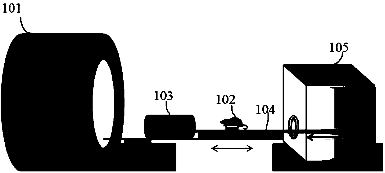
[0025] Please refer to figure 1 , figure 1 It is a structural diagram of a magnetic resonance-compatible autofluorescence tomography molecular imaging device. The optical obscura 105 is connected to the magnetic resonance device 101 through the guide rail 104 to ensure that the posture of the small animal 102 is uniform, and the internal organs of the small animal will not be deformed in the subsequent steps. The optical signal and magnetic resonance are scanned sequentially. The small animal 102 is sent into the optical dark box 105 and the small animal coil 103 through the guide rail 104 .

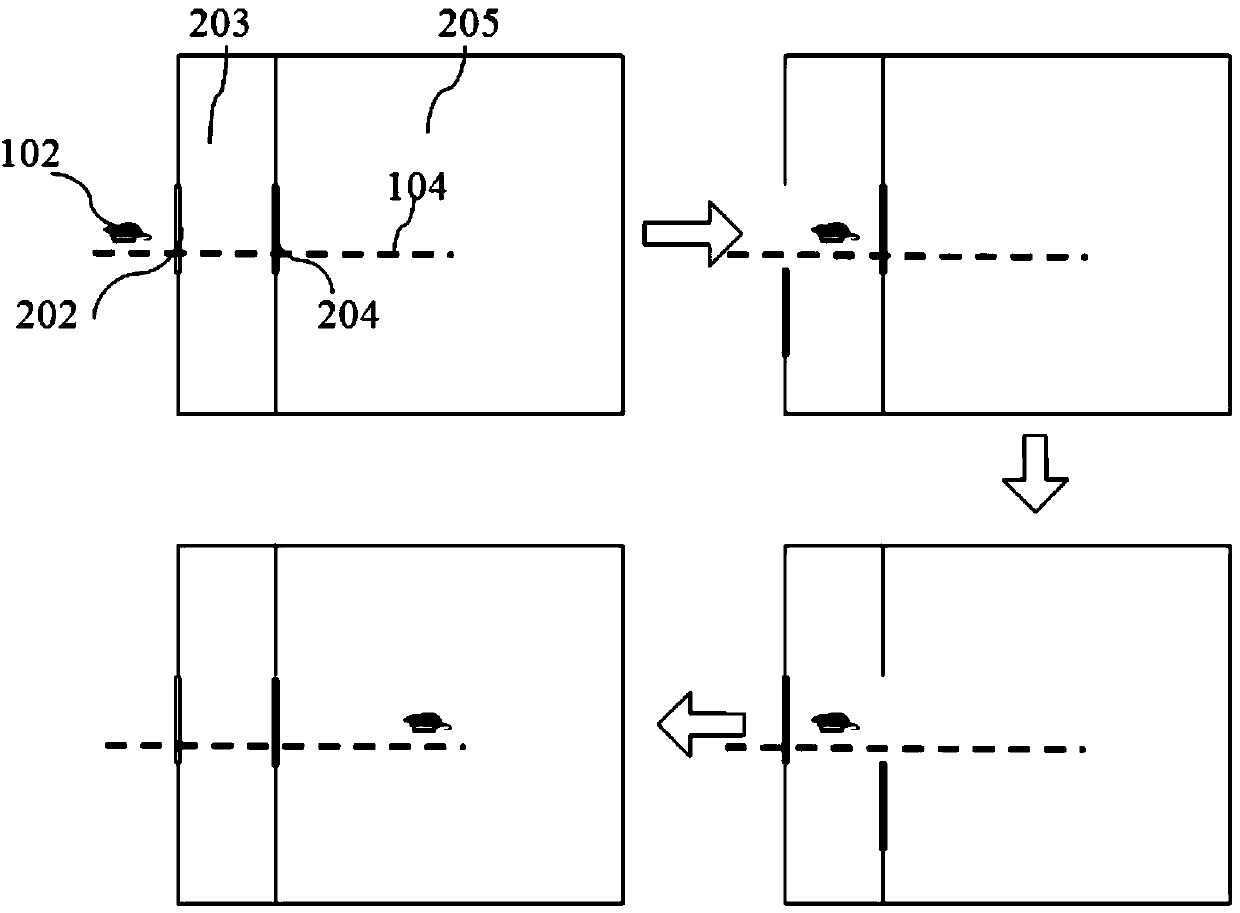
[0026] Please refer to figure 2 , figure 2 It is a schematic diagram of the structure of the shielding shell; it demonstrates how the small animal 102 enters the inner cabin 205 of the optical obscura. The small animal 102 is placed on the guide r...


PUM


Abstract

The invention discloses autofluorescent fault molecular imaging equipment compatible with magnetic resonance. The autofluorescent fault molecular imaging equipment comprises optical imaging equipment and a guide rail; the optical imaging equipment comprises a shielding case and an optical camera obscura, wherein an imaging device is arranged inside the optical camera obscura; the two sides of the guide rail are located in existing medical magnetic resonance equipment and the optical imaging equipment respectively, the guide rail is provided with a carrying platform used for carrying a small animal, and the carrying platform can slide on the guide rail; the shielding case isolates mutual interference between the magnetic resonance equipment and the optical imaging equipment; the carrying platform guarantees that the small animal does not need to be hung and the posture of the small animal does not change in the magnetic resonance imaging process and the optical camera process. A two-sided reflector and rotating wheels are adopted so that the optical imaging equipment can acquire multi-angle multi-spectral autofluorescent data in one plane. High-precision three-dimensional imaging is realized based on the autofluorescent data in combination with structural data provided by the magnetic resonance equipment.

Description

technical field [0001] The present invention relates to a magnetic resonance-compatible autofluorescence tomography molecular imaging device, more specifically, it relates to a multi-spectral and multi-angle autofluorescence tomography device combined with magnetic resonance, which can realize biological internal Noninvasive real-time dynamic in vivo imaging of physiological or pathological processes. Background technique [0002] According to the concept of molecular imaging proposed by Weissleder, all biomedical imaging methods that can characterize and measure biological processes in vivo at the cellular and molecular levels belong to the category of molecular imaging. Currently common molecular imaging modalities mainly include nuclide imaging, such as positron emission tomography, single photon emission computed tomography, magnetic resonance imaging, optical imaging, and ultrasound imaging. Each modal imaging technology has its own advantages and disadvantages, and th...

Claims


Application Information

Patent Timeline
IPC IPC(8): A61B5/00A61B5/055
Inventor 陈多芳张军梁继民陈雪利雷晶薛华丹金征宇田捷
Owner XIDIAN UNIV
Why Eureka
Try Eureka