In-vitro three-dimensional culture method for palatal organ
A three-dimensional culture and organ technology, applied in the field of cell and molecular biology, to achieve the effect of simple operation and low cost
- Summary
- Abstract
- Description
- Claims
- Application Information
AI Technical Summary
Problems solved by technology
Method used
Image
Examples
Embodiment 1
[0028] Example 1: Matrigel three-dimensional culture of palatal plate tissue in vitro
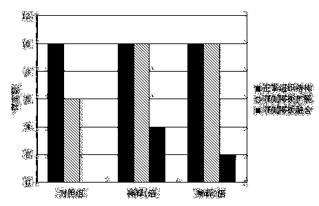
[0029] The middle one-third of the facial tissues (all the tissues from the infraorbital rim to the corner of the mouth, including the palate, the lower part of the nasal septum and surrounding tissues, referred to as palatal organs) were separated from the normal mouse embryonic palate development period (embryonic day 5). Mix with Matrigel Matrigel diluted 1-fold or 2-fold in serum-free DMEM, 10 samples per group, and place in an incubator to solidify into a gel. After 1 hour, DMEM medium containing 10% fetal bovine serum was added for incubation. Unembedded mouse palate tissue was used as the experimental control group (10 samples). All groups were cultured in vitro for 6 days, and fresh culture medium was replaced every two days. After 6 days, the palatal plate tissue of each group was observed, the number of samples with normal palatal plate tissue structure, bilateral palatal plate ...
Embodiment 2
[0035] Example 2: Collagen hydrogel three-dimensional culture of palatal plate tissue in vitro
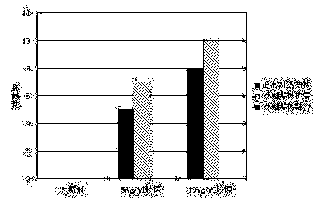
[0036] Separation of tissues from the middle third of the palate during embryonic development of spontaneous cleft palate mice (day 6 of embryonic stage) (all tissues from the infraorbital rim to the corner of the mouth, including the palate, the lower part of the nasal septum and surrounding tissues, referred to as palatal organs) , and then mixed with mouse type II collagen (molecular weight 500kDa) solutions with a concentration of 5 mg / ml and 10 mg / ml respectively, 10 samples in each group, and put them in an incubator to solidify for 40 minutes to form a cylindrical hydrogel with a diameter of 2 cm and a thickness of 1 cm. Glue, and added DMEM medium containing 10% fetal bovine serum for culture. The unembedded palate tissue of mice with spontaneous cleft palate was used as the experimental control group (10 samples). All groups were cultured in vitro for 6 days, and fresh cu...
Embodiment 3
[0041] Example 3: In vitro three-dimensional culture of palatal plate tissue on oxidized hyaluronic acid / collagen composite hydrogel scaffold
[0042] Bovine sodium hyaluronate (molecular weight 1100 kDa) was prepared into a 10 mg / ml aqueous solution, and 20 mM sodium periodate solution was added according to the volume ratio of sodium hyaluronate / sodium periodate at 4:1, and stirred at 25°C in the dark. React for 5 hours, then dialyze, and finally freeze-dry to obtain oxidized hyaluronic acid.
[0043] Prepare 0.9% (w / v) collagen solution, and mix the collagen solution, DMEM medium and NaOH-NaHCO 3 -Hepes buffer is mixed according to the ratio of 7:2:1 (v / v), and then 0.6% (w / v) oxidized hyaluronic acid aqueous solution is mixed according to 10:1 and 30:1 (v / v) (solution: oxidized transparent Aqueous acid solution) was mixed, stirred slowly, and the mixture was placed in a refrigerator at 4 °C for 24 h.
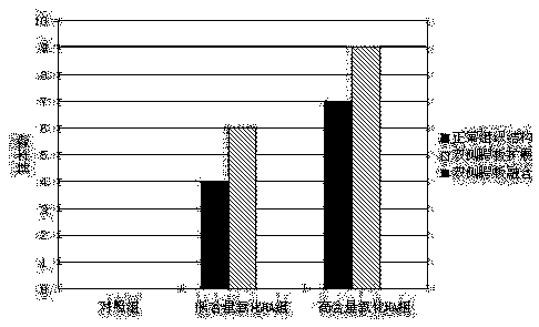
[0044] Separation of the mid-third of the facial tissues of the mouse...
PUM
| Property | Measurement | Unit |
|---|---|---|
| Diameter | aaaaa | aaaaa |
Abstract
Description
Claims
Application Information
 - R&D
- Intellectual Property
- Life Sciences
- Materials
- Tech Scout
- Unparalleled Data Quality
- Higher Quality Content
- 60% Fewer Hallucinations
Browse by: Latest US Patents, China's latest patents, Technical Efficacy Thesaurus, Application Domain, Technology Topic, Popular Technical Reports.
© 2025 PatSnap. All rights reserved.Legal|Privacy policy|Modern Slavery Act Transparency Statement|Sitemap|About US| Contact US: help@patsnap.com


