

Contourlet domain multi-modal medical image fusion method based on statistical modeling
A medical image and statistical modeling technology, applied in the field of medical image processing, can solve problems such as low spatial resolution of fusion images and distortion of spectral information
- Summary
- Abstract
- Description
- Claims
- Application Information
AI Technical Summary
Problems solved by technology
Method used
Image

Examples
Embodiment Construction
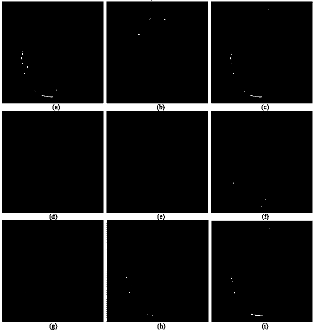
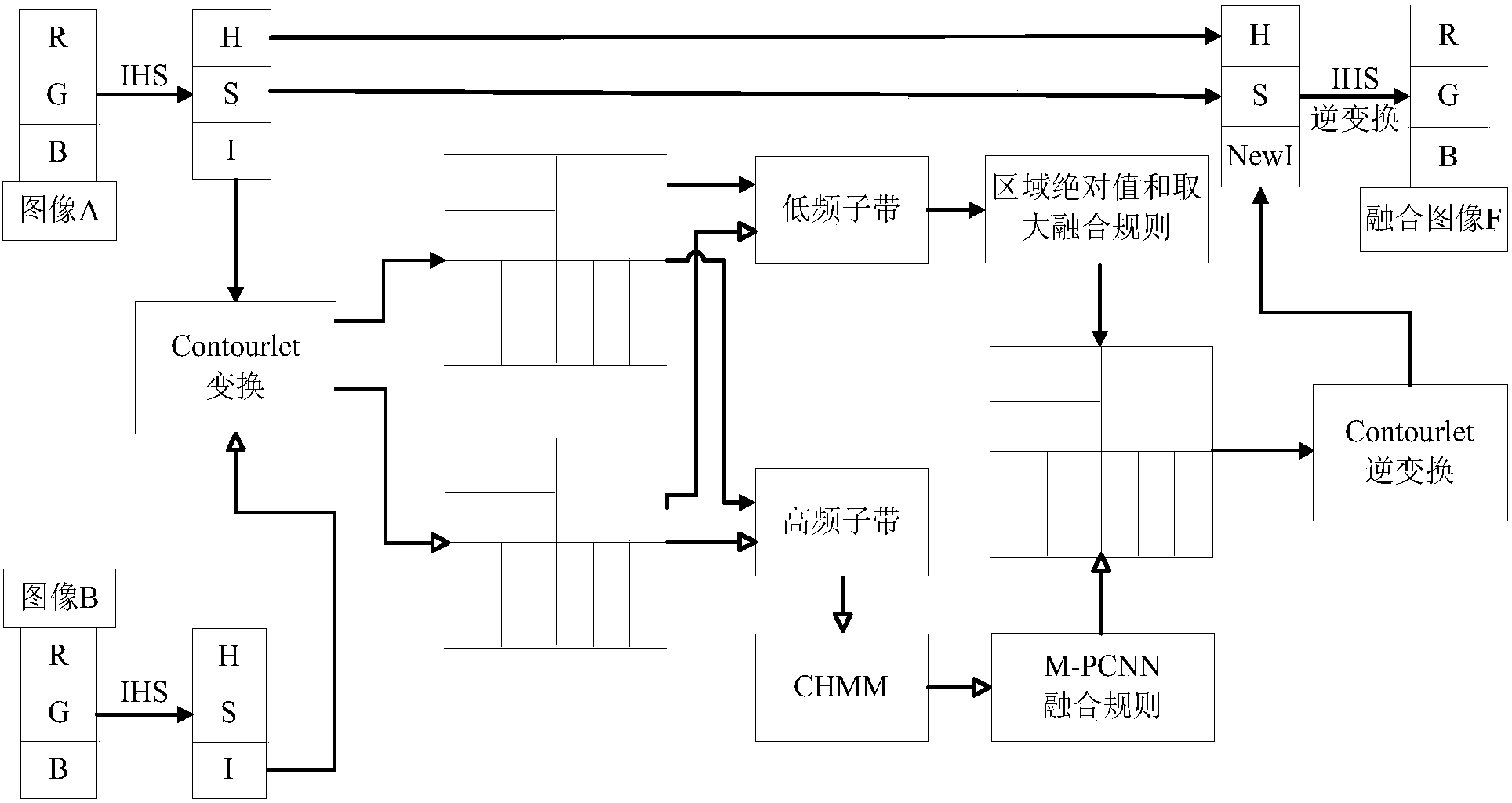
[0058] An embodiment of the present invention (MRI-SPECT medical image) will be described in detail below in conjunction with the accompanying drawings. This embodiment is carried out under the premise of the technical solution of the present invention, as figure 1 As shown, the detailed implementation and specific operation steps are as follows:
[0059] Step 1, perform IHS transformation on the two multimodal medical images to be fused to obtain the corresponding brightness, hue and saturation components;
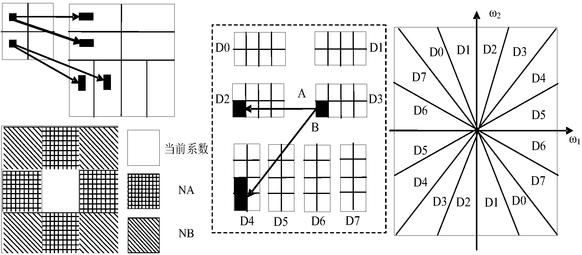
[0060] Step 2, perform Contourlet transform on the luminance component separately, and decompose to obtain high and low frequency subband coefficients of different scales and directions (j=1,2...,J; k=1,2,...,m j ;i,l=1,...,N j ), where “9-7” biorthogonal filter is selected for the scale decomposition LP, “pkva” is selected for the direction filter bank DFB, and the direction decomposition parameter is set to [2,2,3,3], that is, four-scale decomposition is performed, b...
PUM
 Abstract
Description
Claims
Application Information