
Mandibular condyle sagittal fracture reduction forceps

A technology of sagittal fracture and reduction forceps, which is applied in the field of medical devices, can solve the problems of increasing the difficulty of treatment, narrow operating field of view, secondary fractures, etc., and achieve the effect of avoiding peripheral blood vessels and nerves, fully exposing the field of view, and reducing the width

Inactive Publication Date: 2014-09-17
彭国光
View PDF8 Cites 5 Cited by
  • Summary
  • Abstract
  • Description
  • Claims
  • Application Information

AI Technical Summary

Problems solved by technology

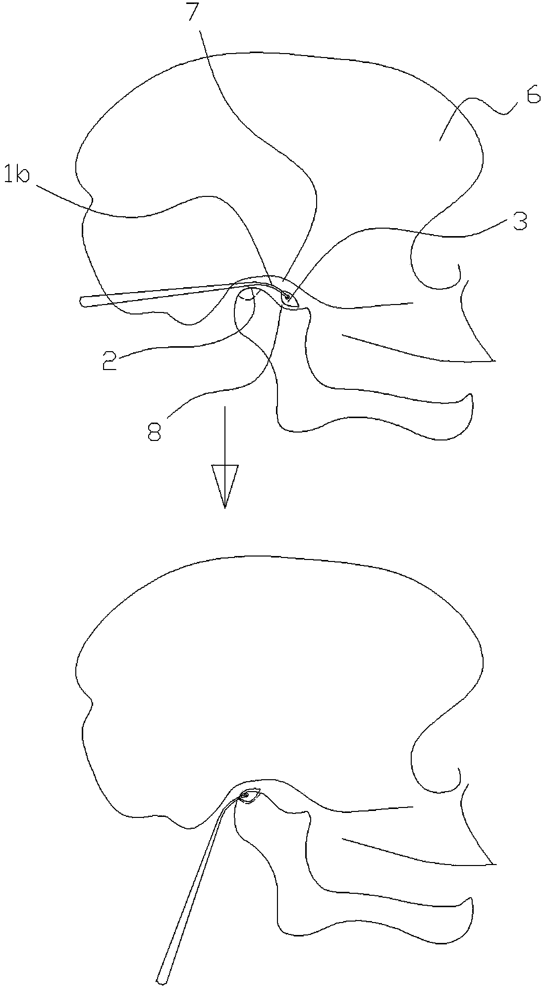
The reasons why sagittal condylar fractures are more difficult to treat than other maxillofacial fractures are: ①The surgical field of view is narrow, located in the deep side of the face, and the position is deep, and doctors cannot anatomically reduce the condyle under direct vision; ②Condylar condyle occurs After the sagittal process fracture, the free medial condylar sagittal fracture fragment shifts inwardly, anteriorly, and inferiorly to the deep side of the face. Due to the lack of suitable surgical instruments, doctors can only use nerve strippers, curved hemostats, Alis Use blind / semi-blind detection methods to find free medial condylar sagittal fracture fragments based on clinical experience; ③ After capturing free medial condylar sagittal fracture fragments, use curved forceps or similar During the reduction process of the instrument, the "secondary fracture" of the fracture fragment is often caused, and more small bone fragments are formed, which increases the difficulty of treatment. Difficult to bend forceps reset, often need to cut off the tendon to completely dissociate the fracture and reset, which means that the blood supply of the condyle is stripped, resulting in the absorption of the condyle after operation; Important vascular and nerve tissues, such as the main trunk and branches of the facial nerve, the posterior facial vein, and the internal maxillary artery, etc., may easily cause damage to the anatomical structure by using inappropriate instruments during operation, resulting in a higher complication rate of condylar sagittal fracture surgery than that of maxillofacial fractures. Other surgical operations, such as internal maxillary artery hemorrhage, facial paralysis, etc.

Method used


Image

  • Mandibular condyle sagittal fracture reduction forceps
  • Mandibular condyle sagittal fracture reduction forceps
  • Mandibular condyle sagittal fracture reduction forceps

Examples


Embodiment Construction

[0013] Now in conjunction with accompanying drawing and embodiment the present invention is described in further detail:

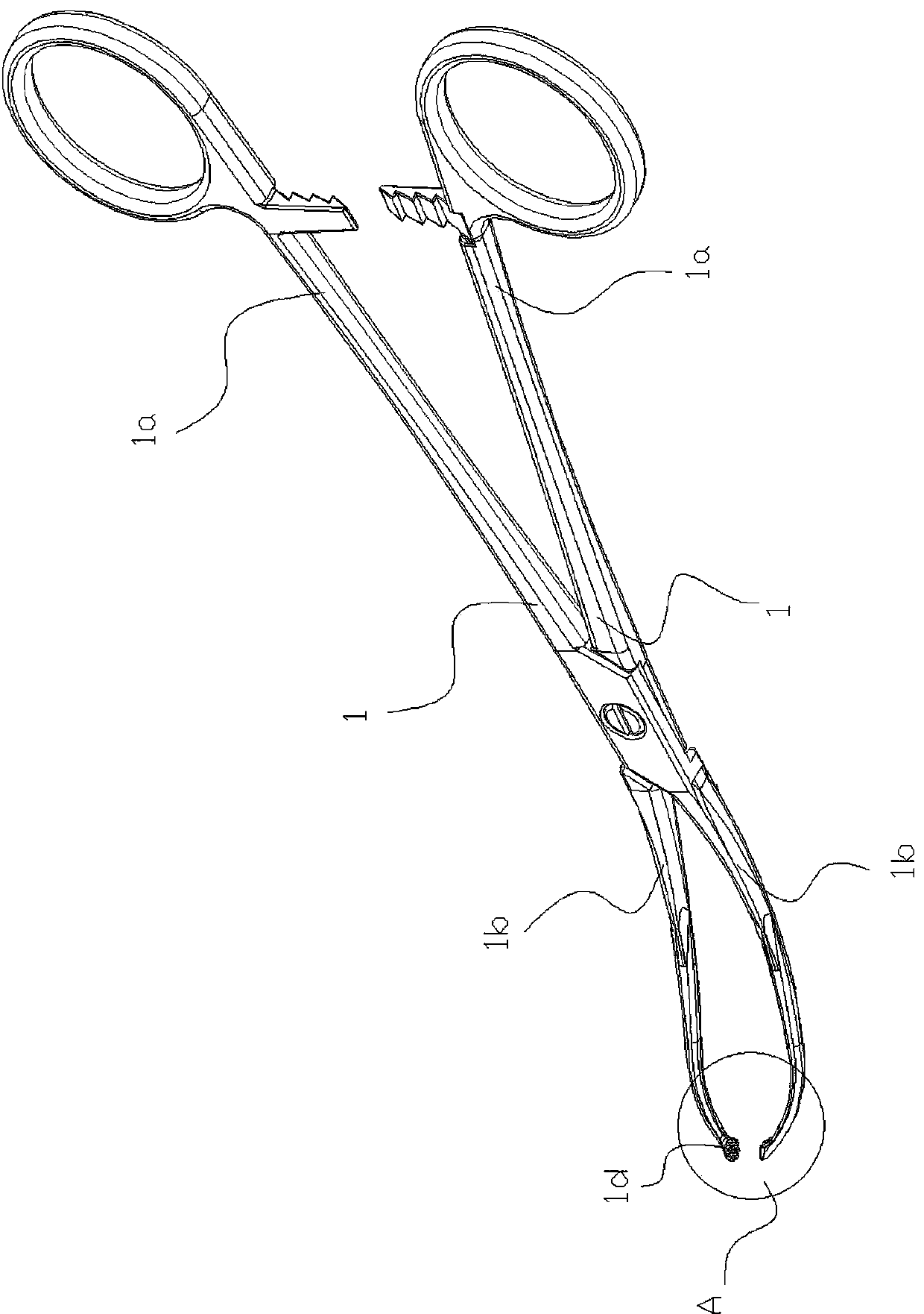
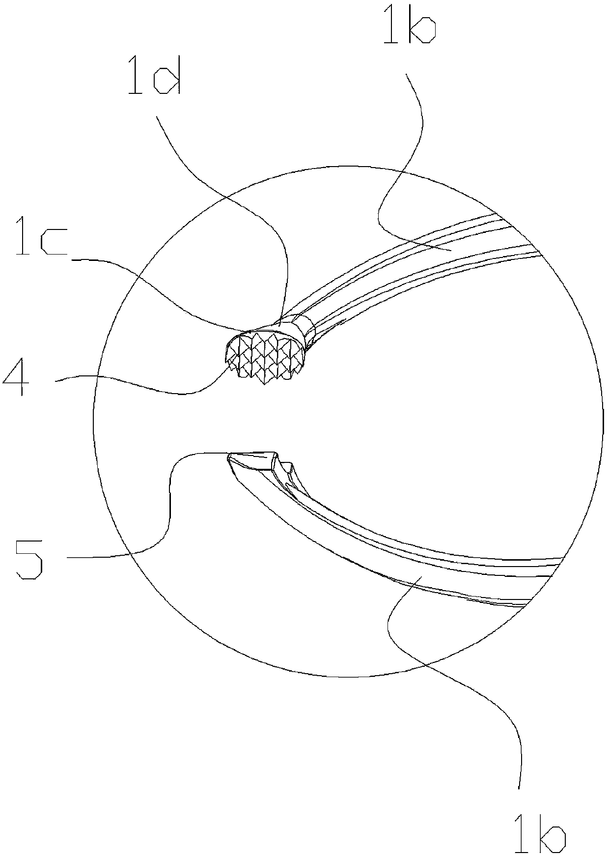
[0014] As shown in the figure, the present invention includes a pair of pliers bodies 1 hinged together. The pliers body 1 includes a handle 1a and a pliers head 1b. 1b starts from the hinge point to the free end, and its cross-sectional size gradually becomes smaller. The free end of the pliers head of one of the pliers body 1b is provided with a chuck 1d whose side is an enlarged plate surface 1c.

[0015] Here, the downward curvature of the forceps head 1 b corresponds to the curvature of the sagittal articular surface 2 of the condyle. The diameter of the downward curved arc of the pliers head 1b is between 50-70mm.

[0016] When clamping, the distance between the outer sides of the two clamp heads 1b is not greater than 10mm.

[0017] In order to prevent the broken bone 3 from slipping off the forceps head 1b, several protrusions 4 are provided on t...


PUM

PropertyMeasurementUnit
Diameteraaaaaaaaaa

Abstract

A pair of mandibular condyle sagittal fracture reduction forceps comprises a pair of sub forceps bodies hinged together. Each sub forceps body comprises a handle and a forceps head. The pair of the forceps is characterized in that the forceps are bent downward while bent inward, the section size of the forceps heads is gradually reduced from the hinge point to the free end, the free end of the forceps head of one sub forceps body is provided with a chuck, and the side face of the chuck is provided with a widening panel. Compared with the prior art, the pair of the mandibular condyle sagittal fracture reduction forceps has the advantages of fully exposing views, avoiding surrounding vessels and nerves, clamping free mandibular condyle sagittal bones, assisting in reduction of the free mandibular condyle sagittal fracture and enabling surfaces of fracture blocks to be uniformly stressed, and the free mandibular condyle sagittal bones can not be broken when clamped.

Description

technical field [0001] The invention relates to a medical device. Background technique [0002] The incidence of sagittal condylar fractures accounts for about 9% to 45% of mandibular fractures. Its treatment has always been the focus and difficulty of oral and maxillofacial trauma. If not treated properly, it is easy to cause traumatic arthritis, ankylosis and other related complications disease. The reasons why sagittal condylar fractures are more difficult to treat than other maxillofacial fractures are: ①The surgical field of view is narrow, located in the deep side of the face, and the position is deep, and doctors cannot anatomically reduce the condyle under direct vision; ②Condylar condyle occurs After the sagittal process fracture, the free medial condylar sagittal fracture fragment shifts inwardly, anteriorly, and inferiorly to the deep side of the face. Due to the lack of suitable surgical instruments, doctors can only use nerve strippers, curved hemostats, Alis ...

Claims


Application Information

Patent Timeline
IPC IPC(8): A61B17/56A61B17/28
CPCA61B17/2812A61B2017/564
Inventor 彭国光
Owner 彭国光
Learn More