
Method and device for correlated micro-imaging of frozen light microscope and frozen electron microscope

A technology of light microscopy and electron microscopy, which is applied to measuring devices, material analysis through optical means, and material analysis using wave/particle radiation. Effects of ice crystal formation, fast and precise realization, fast and precise correlative imaging

Active Publication Date: 2014-12-17
UNIV OF SCI & TECH OF CHINA
View PDF8 Cites 14 Cited by
  • Summary
  • Abstract
  • Description
  • Claims
  • Application Information

AI Technical Summary

Problems solved by technology

The problems of this system are: during the cryo-light microscope imaging process, the sample is exposed to the air, which causes the sample to be easily polluted by ice crystals; the cartridge is small, and only depends on its own gravity to fit the cryo-associated platform, so it is less stable under cryo-conditions Poor, causing the sample to shake a lot during the imaging process; it is only suitable for the Polara electron microscope of FEI Company, and the scope of promotion and use is small
In addition, the cryo-correlation platform developed by the research group of Wolfgang Baumeister of the Max Planck Institute in Germany and the research group of Abraham J.Koster of Leiden University in the Netherlands uses a self-made sample holder to carry the electron microscope sample carrier grid. After the cryo-light microscope imaging is completed, It is necessary to remove the sample grid from the sample holder, transfer it to the sample holder or sample holder of the corresponding electron microscope, and then transfer it to the cryo-electron microscope for imaging, which leads to the need for the sample grid to be transferred multiple times: on the one hand, multiple times The transfer increases the difficulty of correlation; on the other hand, the repeated exposure of frozen samples to air and boiling liquid nitrogen makes the samples unusable due to ice crystal contamination, grid deformation and carbon film breakage

Method used


Image

  • Method and device for correlated micro-imaging of frozen light microscope and frozen electron microscope
  • Method and device for correlated micro-imaging of frozen light microscope and frozen electron microscope
  • Method and device for correlated micro-imaging of frozen light microscope and frozen electron microscope

Examples


Embodiment Construction

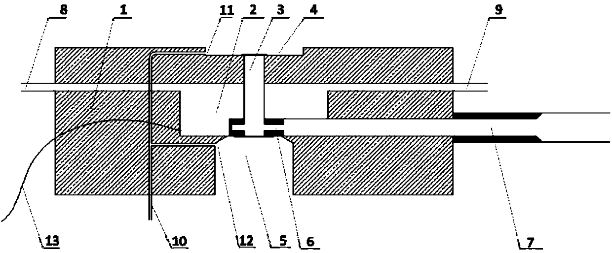
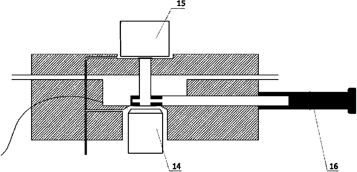
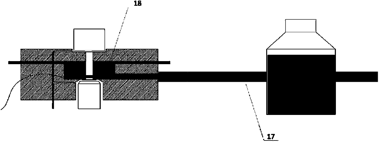
[0020] The invention mainly includes a light microscope cryogenic stage device and a corresponding associated imaging method.

[0021] The optical microscope cryo-stage device of the present invention is a set of carrying devices capable of placing a side-inserted cryo-transmission electron microscope sample rod on an optical microscope platform for cryo-optical microscopic imaging.

[0022] The main technical solution of the present invention is to apply the electron microscope sample rod to the cryo-light microscope imaging. At present, most of the frozen sample rods used in the transmission electron microscope, taking the Gatan 626 sample rod as an example, adopt the sliding cover type sample chamber design, which is about to freeze the sample After being placed in the sample holder, the sample can be sealed in a small space when not imaging, which ensures that the sample is not affected by the external environment, and at the same time maintains the sample in a stable low t...


PUM


Abstract

The invention relates to a method and a device for correlated micro-imaging of a frozen light microscope and a frozen electron microscope, in particular to a method and a device for correlated imaging of a frozen light microscope and a frozen transmission electron microscope, including an optical microscope frozen object stage device used for optics-electron correlated micro-imaging and a corresponding correlated imaging method. By adopting the method and the device, optical micro-imaging at extra-low temperature (below minus 170 degrees) can be achieved, and frozen electron micro-imaging of the same samples in the same areas can also be achieved. The method and the device can be widely used to correlate various types of inverted optical microscopes and most transmission electron microscopes at the market, can achieve correlated imaging fast, conveniently and accurately, prevent ice crystals from forming on surfaces of the samples to the maximum extent, avoid problems of grid deformation and carbon film fracture, and can be widely applied to related research of cytobiology, neurobiology, biological medicines, etc.

Description

technical field [0001] The present invention relates to equipment and methods for microscopic imaging, in particular to the associated imaging of optical microscopy and electron microscopy, especially the associated imaging of cryo-optic microscopy and cryo-transmission electron microscopy, including optical-electron microscopy associated imaging. Microscope cryo-stage setup and corresponding associated imaging methods. Background technique [0002] Cryo-photocorrelation microscopy imaging: a method that can perform optical microscopic detection of samples labeled with optical probes at extremely low temperatures (below -170 ° C, capable of maintaining the fine structure information of samples in close to physiological states), and A technique that enables high-resolution electron microscopic imaging detection of the same area of ​​the sample. Cryo-photonic correlation microscopy combines the advantages of modern fluorescence microscopy and cryo-electron microscopy to make ...

Claims


Application Information

Patent Timeline
IPC IPC(8): G01N21/01G01N23/00
Inventor 毕国强周正洪何继中陶长路徐程刘云涛
Owner UNIV OF SCI & TECH OF CHINA
Learn More