
Method of intracellular infectious agent detection in sperm cells

A technology of sperm cells and cells, applied in the field of detection of intracellular infectious agents in sperm cells

Inactive Publication Date: 2015-01-21
瓦西利奥斯·齐列瓦可斯 +1
View PDF4 Cites 3 Cited by
  • Summary
  • Abstract
  • Description
  • Claims
  • Application Information

AI Technical Summary

Problems solved by technology

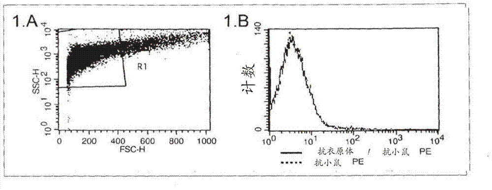
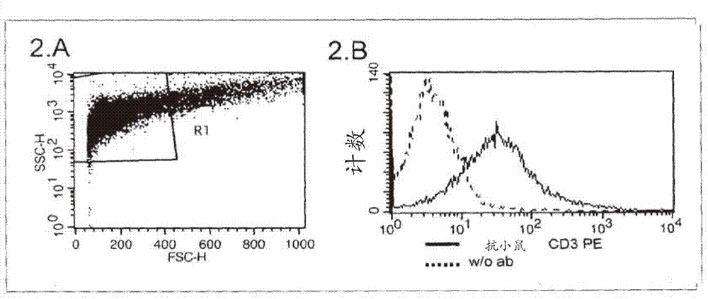
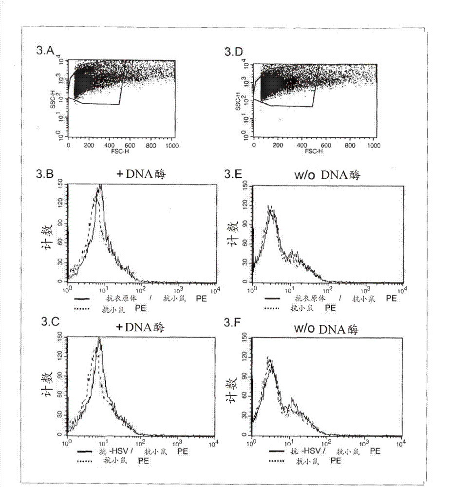
[0035] The above results show that the method proposed by Stewart et al. in patent application US 2006 / 0099661 A1 for the detection of cellular endoplasma in sperm cells does not provide a solution to the problems indicated by the present invention

Method used


Image

  • Method of intracellular infectious agent detection in sperm cells
  • Method of intracellular infectious agent detection in sperm cells
  • Method of intracellular infectious agent detection in sperm cells

Examples


Embodiment Construction

[0059] An example of an embodiment of the invention follows:

[0060] 1. fixation of sperm cells including sperm

[0061] After semen collection and liquefaction, spermatozoa were centrifuged and fixed with 4% paraformaldehyde (PFA) for 30 min at 4°C. PFA works by cross-linking proteins, thereby inactivating pathogens, and immobilizing putative autoantibodies that have been attached to sperm, making them detectable, if that is desired. In some rare cases, some epitopes may be altered or destroyed so that they cannot be detected by certain antibodies. In this case, surface staining can be performed prior to fixation. Furthermore, PFA fixation preserves the physical properties of the cells: namely, after fixation, these cells exhibit the same scattering properties as those performed by flow cytometry during the analysis. Furthermore, PFA fixation allows the subsequent application of an extracellular or intracellular staining procedure.

[0062] 2. Important notice: Befor...


PUM


Abstract

The present invention describes a method for investigating the presence of chlamydia, viruses and other infectious agents in spermatozoa, with the use of immunofluorescence in combination with flow cytometry. This method is used for the intracellular detection of microorgaqnisms within sperm cells through the use of a DNA easing procedure, as well as for the detection of microorganisms adherent to the surface of spermatozoa.

Description

technical field [0001] The present invention describes a method for subjecting semen, i.e. a population of sperm cells, to analysis for the detection of intracellular viruses, chlamydia, parasites and other micro-organisms inside the sperm in an attempt to study and determine the causes of subfertility and infertility etiology, and prevention of congenital infections. The entire procedure is done using a specific process for alleviating the DNA structure, targeting the microorganisms with antibodies and evaluating the results with flow cytometry. Background technique [0002] Subfertility is an increasingly common problem affecting many families. To address this problem, a number of methods have been developed, including in vitro fertilization (IVF). [0003] Blocked fallopian tubes, azoospermia and menopause are undisputed causes of subfertility. Oligospermia and premenopause, the early stages of azoospermia and menopause, are also of interest because they are important ...

Claims


Application Information

Patent Timeline
Patent Type & Authority Applications(China)
IPC IPC(8): G01N33/569
CPCG01N33/56927G01N33/56983C12Q1/70G01N33/533G01N33/569G01N33/56905G01N33/56988G01N33/56994G01N2800/367C12Q1/04C12Q1/34G01N33/577
Inventor 瓦西利奥斯·齐列瓦可斯阿格勒斯·格瑞扎培斯
Owner 瓦西利奥斯·齐列瓦可斯
Learn More