CUDA-based dental three-dimensional CT image processing method

A technology of three-dimensional image and processing method, applied in image data processing, 3D image processing, instruments, etc., can solve problems such as difficult system, slow processing speed, and high cost, and achieve high computing speed, cost reduction, and fast three-dimensional display Effect

Inactive Publication Date: 2015-02-11
NANJING PERLOVE RADIAL VIDEO EQUIP
View PDF2 Cites 0 Cited by
  • Summary
  • Abstract
  • Description
  • Claims
  • Application Information

AI Technical Summary

Problems solved by technology

[0002] The auxiliary role of medical imaging in doctor's diagnosis is becoming more and more obvious. In order to improve the accuracy and scientificity of medical diagnosis and treatment planning, it is necessary to transform the sequence of two-dimensional tomographic images into three-dimensional images with intuitive three-dimensional effects. However, the current three-dimensional medical Auxiliary diagnostic systems all require a workstation-level operating platform to basically meet the requirements for processing a large amount of image data. Due to the price problem, this system is difficult to promote. At present, GPUs specially used for image processing are more and more widely used in computer graphics and images. , a GPU with ultra-large-scale data processing and computing capabilities, has the incomparable speed advantage of a CPU, and realizes large-scale data processing on consumer-grade GPU hardware. The CUDA programming language is a C language environment for general-purpose computing GPUs, and CUDA programming is applied GPU general-purpose computing can be easily realized. The current general-purpose volume rendering technology is generally based on the traditional CPU for calculation, and the processing speed is relatively slow. Generally, it needs to be run on a parallel graphics workstation, and the cost is relatively high.

Method used


Image

  • CUDA-based dental three-dimensional CT image processing method

Examples


Embodiment Construction

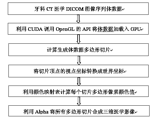
[0015] With reference to accompanying drawing, the dental CT three-dimensional image processing method based on CUDA is characterized in that: the method may further comprise the steps:

[0016] The first step is to read in the DICOM image sequence file and save it to the system memory in the form of volume data;

[0017] Volume data is composed of sequential two-dimensional medical DICOM image sequences. The image resolution, layer spacing and image pixel information of these medical images are read into the system memory. The volume data is first preprocessed, such as denoising with image filters, And by interpolating layer spacing data to get a more detailed effect;

[0018] The second step is to use CUDA to call the 3D graphics library programming extension API of OpenGL to load the volume data in the system memory into the GPU memory;

[0019] The third step is to calculate and generate polygonal slices, which are divided into the following aspects:

[0020] 1. Calculat...


PUM


Abstract

The invention discloses a CUDA-based dental three-dimensional CT image processing method and belongs to the technical field of medical image processing. The method is characterized by comprising the following steps: a first step of reading in a DICOM image sequence file and storing the DICOM image sequence file in an internal memory of a system in a form of volume data; a second step of calling a three-dimensional graphic library programming extension port function API of OpenGL by using CUDA and loading the volume data in the internal memory of the system into a GPU video memory; a third step of calculating and generating polygonal segments in the following processes: (1) calculating a coordinate of each apex of the volume data in a viewpoint coordinate system; (2), calculating the number of the polygonal segments and calculating a world coordinate of each apex of each polygonal segment; (3) integrating each polygonal segment into a three-dimensional medical image. The method has the advantages that the calculation rate is high, the quick three-dimensional display of the medical image on a general PC (Personal Computer) at a common consumption level can be implemented without a graphic workstation, and the cost is greatly lowered.

Description

technical field [0001] The invention belongs to the technical field of medical image processing. Background technique [0002] The auxiliary role of medical imaging in doctor's diagnosis is becoming more and more obvious. In order to improve the accuracy and scientificity of medical diagnosis and treatment planning, it is necessary to transform the sequence of two-dimensional tomographic images into three-dimensional images with intuitive three-dimensional effects. However, the current three-dimensional medical Auxiliary diagnostic systems all require a workstation-level operating platform to basically meet the requirements for processing a large amount of image data. Due to the price problem, this system is difficult to promote. At present, GPUs specially used for image processing are more and more widely used in computer graphics and images. , a GPU with ultra-large-scale data processing and computing capabilities, has an incomparable speed advantage of a CPU, and realize...

Claims


Application Information

Patent Timeline
Patent Type & Authority Applications(China)
IPC IPC(8): G06T15/00
CPCG06T15/005
Inventor 李劲生吴林成
Owner NANJING PERLOVE RADIAL VIDEO EQUIP
Why Eureka
Try Eureka