
Method for making aortic endothelial cell injury model by utilizing mice and detection method

A technology of endothelial cells and injury models, which can be used in measurement devices, veterinary surgery, and veterinary instruments to solve problems such as atherosclerosis

Inactive Publication Date: 2015-03-11
THE FIRST AFFILIATED HOSPITAL OF GUANGZHOU MEDICAL UNIV (GUANGZHOU RESPIRATORY CENT)
View PDF0 Cites 2 Cited by
  • Summary
  • Abstract
  • Description
  • Claims
  • Application Information

AI Technical Summary

Problems solved by technology

[0007] Mice are cheap and easy to operate. Although their fat metabolism process is different from that of humans and their blood lipids are not high, so they are not prone to atherosclerosis, but the apolipoprotein E gene knockout (ApoE - / - ) successful preparation of the mouse model, making this mouse prone to atherosclerosis, and the pathological process is similar to that of humans

Method used


Image

  • Method for making aortic endothelial cell injury model by utilizing mice and detection method

Examples


Embodiment Construction

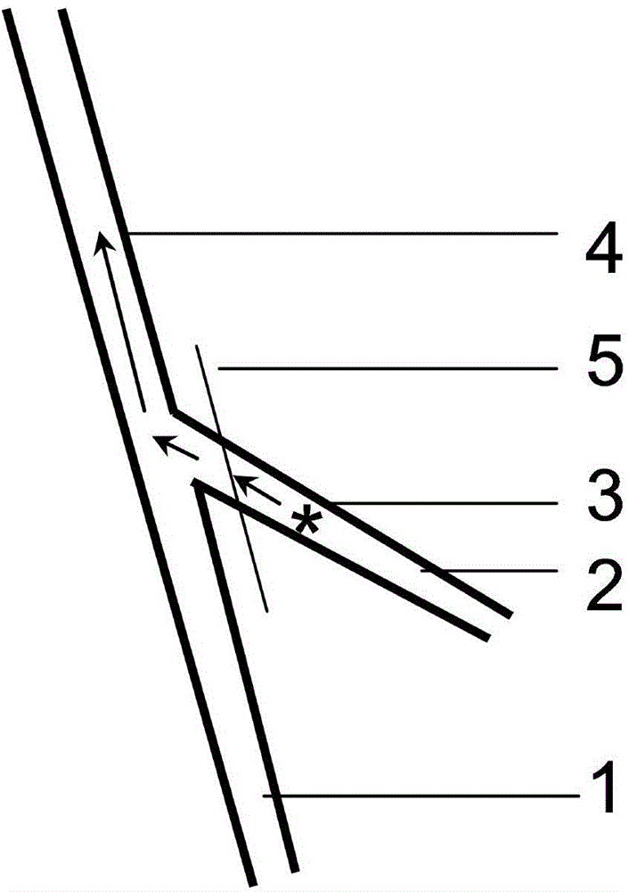
[0032] refer to figure 1 The method for utilizing mouse to make arterial endothelial cell injury model shown, its method step is:

[0033] (1) Preoperative preparation: After general anesthesia by intraperitoneal injection of pentobarbital sodium (120 mg / kg), the mice were placed supine on the operating table, and the limbs were fixed with adhesive tape.

[0034] (2) Surgical procedure:

[0035] A. Shaving and disinfection: shave one side (left side for right-handed patients) and sterilize with iodine in the groin area. The other side did not move, used as a control.

[0036] B. Separate the femoral artery and its branches: cut the skin longitudinally in the middle of the groin, and separate the main trunk of the femoral artery (marked 1 in the figure). Two surgical sutures were passed under the proximal and distal femoral arteries and secured with tape to temporarily control blood flow. Separate the thicker branch of the artery in the middle of the trunk - the muscular br...


PUM

PropertyMeasurementUnit
diameteraaaaaaaaaa

Abstract

The invention discloses a method for making an aortic endothelial cell injury model by utilizing mice and a detection method. The method for making the aortic endothelial cell injury model disclosed by the invention comprises the following steps of: (1), cutting femoral artery surface skin of mice, and separating femoral artery from a femoral artery branch; (2), shearing a small hole on the femoral artery branch, and inserting a spiral stainless steel wire in a femoral artery trunk through the sheared hole of the femoral artery branch; (3), drawing out the spiral stainless steel wire after staying for 1-3 min, ligating the femoral artery branch, and suturing skin; and (4), making an aortic section slice from injured femoral artery after 3-5 weeks, and then, making a specimen. According to the invention, mice can be used as experimental objects for mechanical injury of aortic endothelial cells; in addition, the purpose of researching the reaction degree of the aortic endothelial cells to injury can be achieved with the help of the detection process of the aortic endothelial cells.

Description

technical field [0001] The invention relates to injury of arterial endothelial cells during animal experiments, in particular to a method for making a mechanical injury model of mouse femoral artery endothelial cells and a detection method thereof. The arterial endothelial cell injury method is used for the study of arterial endothelial cells and related diseases, such as the mechanism and therapeutic effect of atherosclerosis. Background technique [0002] The anatomy of a large or medium-sized artery wall is divided into three layers: arterial intima, media, and adventitia. Among them, the arterial intima is located on the inner side of the blood vessel cavity and directly contacts the blood. It is composed of a layer of arterial endothelial cells, which protects the arterial wall from harmful components in the blood. In addition, it secretes some biological substances to regulate the contraction and relaxation of arteries. , so as to maintain the normal function of the c...

Claims


Application Information

Patent Timeline
Patent Type & Authority Applications(China)
IPC IPC(8): A61D1/00G01N33/483
CPCA61D1/00
Inventor 李君沈粤春陆东风李新春
Owner THE FIRST AFFILIATED HOSPITAL OF GUANGZHOU MEDICAL UNIV (GUANGZHOU RESPIRATORY CENT)
Learn More