Use of antigen group in preparation of disease diagnosis kit and kit

A kit and antigen technology, applied in the field of biomedicine, can solve the problems of incompatibility, poor sensitivity and specificity, no simple early diagnosis and prediction, etc., and achieve the effect of broad application prospects.

Active Publication Date: 2015-03-18
CHINA JAPAN FRIENDSHIP HOSPITAL
View PDF1 Cites 3 Cited by
  • Summary
  • Abstract
  • Description
  • Claims
  • Application Information

AI Technical Summary

Problems solved by technology

At present, the clinical screening methods for tumors, such as tumor markers, CT, MRI, PET-CT, etc., are either poor in sensitivity and specificity, unable to achieve early diagnosis, or expensive, and not suitable for screening. means of investigation
Therefore, there is no simple, practical, sensitive and specific index that can diagnose and predict the occurrence of tumors in the early stage.

Method used


Image

  • Use of antigen group in preparation of disease diagnosis kit and kit
  • Use of antigen group in preparation of disease diagnosis kit and kit
  • Use of antigen group in preparation of disease diagnosis kit and kit

Examples


Embodiment 1

[0082] (A) Preparation of Tif1-γ antigen:

[0083] (1) Total RNA was extracted from K562 cells in logarithmic phase (purchased from ATCC, Cat. No. CCL-243) by classical Trizol method, and then reverse-transcribed into cDNA by Promega reverse transcription kit.

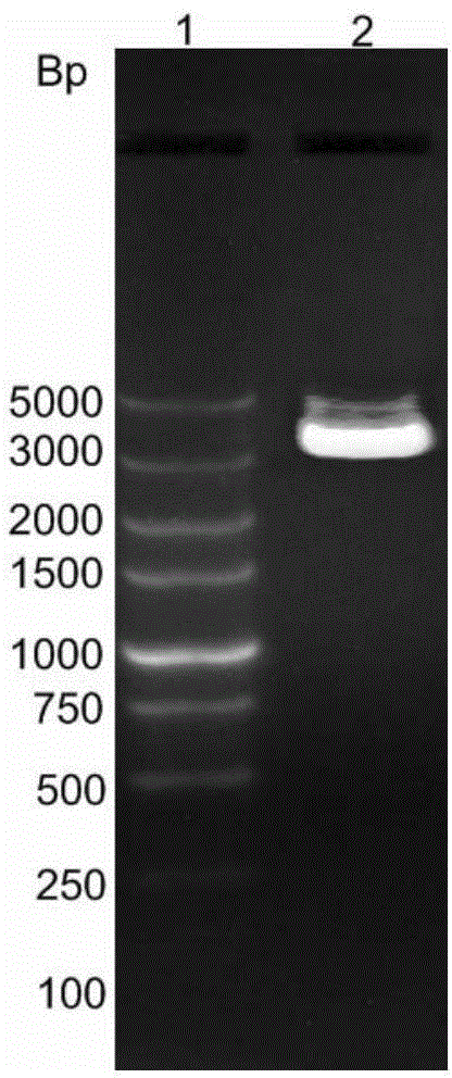
[0084] (2) Using the above cDNA as an amplification template, using Tif1-γ-T1 (5'-TAGCGATCGCCATGGCGGAAAACAAAGG-3', SEQ ID NO.4) and Tif1-γ-T2 (5'-CGCACGCGTCTTTATATGTACTGGTCTCTCAT-3', SEQ ID The primer pair composed of NO.5) was used to amplify the Tif1-γ gene by PCR. The reaction conditions were: the template was denatured at 98°C for 2 minutes, and then reacted according to the following parameters for a total of 35 cycles: 94°C pre-denaturation for 2 minutes, 94°C denaturation for 45s, Anneal at 54°C for 30s, extend at 72°C for 140s, 34 cycles; last cycle at 72°C, extend for 5min, PCR reaction products were identified by 10g / L agarose gel electrophoresis, the identification results are as follows figure 1 shown.

...

Embodiment 2

[0118] The disease detection kit 2 was prepared according to the same method as in Example 1, except that the kit also included: phosphate buffered saline with 20 volume % goat serum used as a sample buffer, 0.5 volume % goat serum used as a washing buffer % Tween 20 in phosphate buffer, 5-bromo-4-chloro-3-indolyl-phosphate and tetrazolium nitro blue (purchased from Millipore; Cat. No. 203790) as substrate, and Incubation plates purchased from Bio-rad.


PUM


Abstract

The invention provides a disease detection kit, the kit includes human IgG (Intravenous Gamma Globulin), Tif1-gamma (transcription intermediary factor 1-gamma) antigen, NXP2 antigen carrier, alkaline phosphatase labeled goat anti-human IgG as second antibody, an anti Tif1-gamma antibody positive comparison product as a contrast, and an anti-NXP2 antibody positive comparison product; and the human IgG, the Tif1-gamma antigen and the NXP2 antigen respectively coats different regions of the carrier. The invention also provides use of the antigen group in preparation of the disease diagnosis kit, the antigen group includes the Tif1-gamma antigen and the NXP2 antigen, and the disease is dermatomyositis merged tumor. The disease detection kit can highly specifically diagnose the dermatomyositis merged tumor by detection of the presence of anti Tif1-gamma antibody and anti-NXP2 antibody, can be used for early diagnosis of the dermatomyositis merged tumor, and has broad application prospects.

Description

technical field [0001] The invention relates to the field of biomedicine, in particular to a kit for detecting a disease and the use of an antigen group in preparing a kit for diagnosing a disease. Background technique [0002] Idiopathic inflammatory myopathy (Idiopathic inflammatory myopathy, IIM) is a group of acquired heterogeneous diseases with skeletal muscle involvement as the main manifestation, mainly including polymyositis (polymyositis, PM), dermatomyositis ( There are 3 types of dermatomyositis (DM) and inclusion body myositis (IBM). It is not uncommon to observe cases of IIM with cancer-associated myositis (CAM) in clinical practice. Previous studies at home and abroad have shown that the risk of IIM patients with cancer is 6%-70%. Among them, the incidence of DM with cancer Significantly higher than PM. The treatment of IIM combined with tumors is more difficult and the mortality rate is high, which is an important factor affecting the prognosis of patients. ...

Claims


Application Information

Patent Timeline
IPC IPC(8): G01N33/544
CPCG01N33/6893
Inventor 卢昕王国春杨阚波彭清林舒晓明
Owner CHINA JAPAN FRIENDSHIP HOSPITAL
Why Eureka
Try Eureka