
Gene joint detection method and kit

A combined detection and kit technology, applied in the field of molecular biology, can solve the problems of patient payment, a large number of clinical samples, high detection costs, etc., and achieve the effect of strong selectivity, strong specificity, and low sample consumption

Active Publication Date: 2015-04-01
南宁维尔凯生物科技有限公司
View PDF0 Cites 4 Cited by
  • Summary
  • Abstract
  • Description
  • Claims
  • Application Information

AI Technical Summary

Problems solved by technology

In lung cancer patients, there are 16 most common mutations of KRAS, PIK3CA, BRAF and EGFR genes. If the method of fluorescent quantitative PCR is used for detection, a large number of clinical samples are required, which not only increases the difficulty of clinical sampling, but also makes the patient Pay higher testing fees

Method used


Image

  • Gene joint detection method and kit
  • Gene joint detection method and kit
  • Gene joint detection method and kit

Examples


Embodiment 1

[0044] Take 2 μL of the DNA in Table 2 and put it in a PCR tube as a template, place it on a PCR instrument, set the temperature at 95°C, maintain it for 5min, and then cool it down to 25°C. Add 1.5 μL MLPA Buffer and 1.5 μL Probemix (the Probemix contains 34 probes in Table 1) to the denatured template, and run the hybridization program on the PCR machine: 95°C for 1min; 60°C for 16h. After the hybridization reaction, add 15 μL of ultrapure water and 0.1 μL of ligase to each PCR tube, and run the ligation program on the PCR instrument: 54°C for 20 minutes; 95°C for 10 minutes; cool to 20°C. PCR amplification was performed after the ligation reaction was completed. Add 1.0 μL of PCR universal primers, 1.0 μL of dNTPs, and 0.5 μL of Taq enzyme to each PCR tube, and run the PCR program on the PCR machine:

[0045]

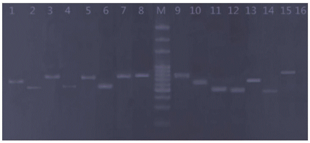
[0046] Detection of amplified nucleic acid fragments by polyacrylamide electrophoresis, see figure 1 . Each mutation point is accurately detected, and the leng...

Embodiment 2

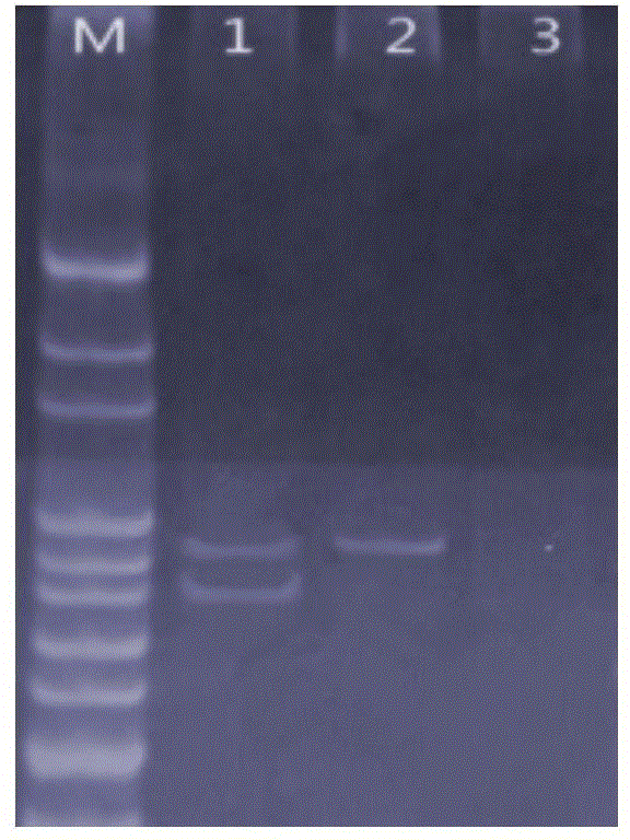
[0050] Take 2 μL of the DNA in Table 3 as a template in a PCR tube, and the rest of the inspection operations are the same as in Example 1, and the amplified nucleic acid fragments are detected by polyacrylamide electrophoresis, see figure 2 . When the DNA extracted from the blood of healthy people was used as the sample, two bands with a length of 186bp and 194bp were amplified by PCR; however, when the genomic DNA of HCC827 cells with E746-A750 deletion was used as a sample, only a band with a length of 194bp was amplified. a strip.

[0051] Table 3: Template DNA of Example 2

[0052]

Embodiment 3

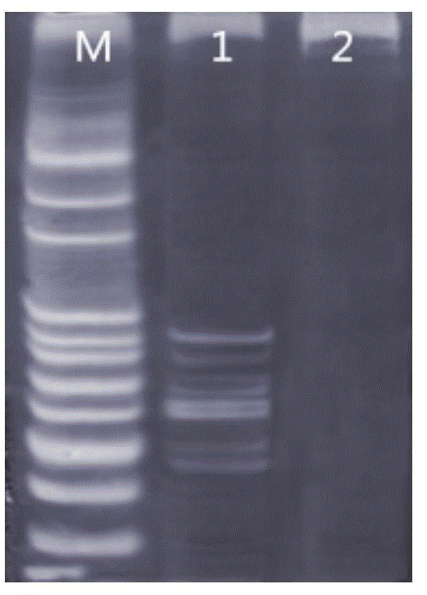
[0054] Take 2 μL of the DNA in Table 4 as a template in a PCR tube, and the rest of the inspection operations are the same as in Example 1, and the amplified nucleic acid fragments are detected by polyacrylamide electrophoresis, see image 3 . When multiple gene mutations exist in the sample at the same time, the test method can detect the mutated genes at the same time.

[0055] Table 4: Template DNA of Example 3

[0056]


PUM


Abstract

The invention relates to a joint detection method for gene mutation. The joint detection method is characterized in that 16 gene mutations, including KRAS (Kirsten rat sarcoma viral oncogene homolog), PIK3CA (phosphatidylinositol-4, 5-bisphosphate 3-kinase, catalytic subunit alpha), BRAF (B-Raf proto-oncogene, serine / threonine kinase) and EGFR (Epidermal Growth Factor Receptor) genes can be detected synchronously by using 20-50ng of DNA as a sample. The method comprises the steps of designing a specific probe; building a reaction system for an MLPA (Multiplex Ligation Dependent Probe Amplification) mutant gene sequences; largely amplifying the targeted gene sequences by PCR (Polymerase Chain Reaction). The joint detection method for gene mutation has the advantages that the specificity is high, and 10-100ng of wild type DNA do not generate nonspecific interference signals; the selectivity is outstanding, and 0.1 to 1.0% of mutation can be detected under the background of 99-99.9% of wild type DNA.

Description

technical field [0001] The invention relates to the field of molecular biology, in particular to a method for joint detection of gene mutations, in particular to a method for simultaneously detecting a total of 16 mutations of KRAS, PIK3CA, BRAF and EGFR genes using the MLPA method as a technical platform. Background technique [0002] RAS genes include KRAS, HRAS and NRAS. The KRAS gene is mainly related to lung cancer. It is located on chromosome 12 and encodes the P21 protein. Regulates cell growth and differentiation. When exons 2 and 3 of the KRAS gene are mutated, the mutated KRAS gene can continuously activate the RAF-MEK-ERK-MAPK signaling pathway, leading to malignant cell proliferation, differentiation, and regulation disorder. A high mutation rate of the KRAS gene has been found in leukemia, lung cancer, colorectal cancer, and pancreatic cancer. [0003] Phosphatidylinositol-3-kinase (PI3K) is an important signaling pathway molecule, which consists of a catalyti...

Claims


Application Information

Patent Timeline
IPC IPC(8): C12Q1/68C12N15/11
CPCC12Q1/6844C12Q1/6886C12Q2600/156C12Q2531/113C12Q2537/143C12Q2561/125
Inventor 唐涛周细武
Owner 南宁维尔凯生物科技有限公司
Learn More