Hepatocarcinoma marker melanoma cell-adhesion molecule and application thereof
A technology for melanoma and liver cancer, which is applied in the field of markers for the diagnosis of liver cancer and its application, and can solve the problems of increased negative ratio of AFP, unsatisfactory sensitivity and specificity, etc.
- Summary
- Abstract
- Description
- Claims
- Application Information
AI Technical Summary
Problems solved by technology
Method used
Image
Examples
Embodiment 1
[0156] Detection of MCAM expression in human liver cancer and normal liver tissues by tissue microarray (TMA) analysis
[0157] 418 liver cancer tissue samples and 24 normal liver tissue samples were stained and analyzed by tissue microarray (TMA). Such as Figure 3A As shown, 304 cases of immunohistochemical membrane staining were positive in 418 cases of liver cancer tissues, the ratio was about 72.7%. However, none of the 24 cases of normal liver tissue stained positively.
[0158] Data analysis showed that MCAM protein expression was significantly stronger in membrane staining in liver cancer tissues than in normal liver tissues.
Embodiment 2
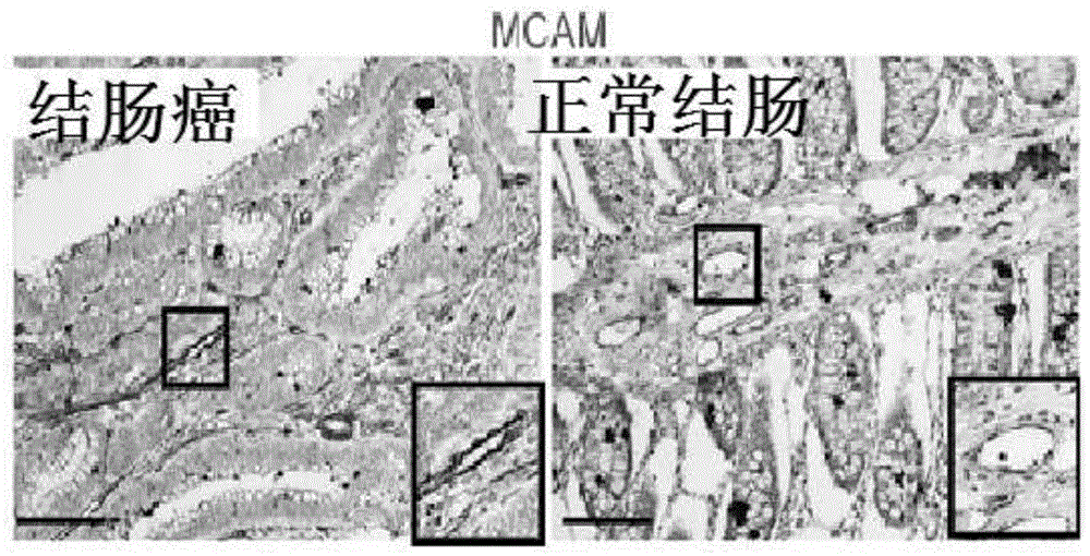
[0160] Immunohistochemical Analysis of MCAM Expression in Human Colon and Breast Cancer Tissues
[0161] IHC staining and analysis were performed on 5 normal and 24 colon cancer tissue samples, and the staining of the corresponding tissue cells was counted. Such as Figure 3G As shown, no statistical difference was found between normal tissue and colorectal cancer tissue samples (p<0.01). In addition, it can be seen that MCAM is mostly located on the surface of acini or blood vessels, and rarely seen on the surface of colon tissue cells.
[0162] Similarly, 5 cases of normal and 24 cases of breast cancer tissue samples were stained and analyzed by IHC, and the staining conditions of corresponding tissue cells were counted. Such as Figure 3H As shown, no statistical difference was found between normal tissue and breast cancer tissue samples (p<0.01). In addition, it can be seen that MCAM is mostly located on the surface of acini or blood vessels, and rarely seen on the sur...
Embodiment 3
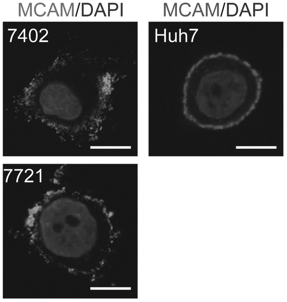
[0165] A group of liver cancer cell lines SMMC-7721, Bel-7402 and Huh7 were selected to observe the membrane localization of MCAM by immunofluorescence. Such as Figure 1B As shown, it can be seen that MCAM is clearly located on the cell membrane.
[0166] Data analysis revealed that MCAM protein is a liver cancer-specific marker located on the cell membrane.
PUM
 Abstract
Description
Claims
Application Information
 - R&D
- Intellectual Property
- Life Sciences
- Materials
- Tech Scout
- Unparalleled Data Quality
- Higher Quality Content
- 60% Fewer Hallucinations
Browse by: Latest US Patents, China's latest patents, Technical Efficacy Thesaurus, Application Domain, Technology Topic, Popular Technical Reports.
© 2025 PatSnap. All rights reserved.Legal|Privacy policy|Modern Slavery Act Transparency Statement|Sitemap|About US| Contact US: help@patsnap.com


