Digital X-ray image contrast enhancement processing method

A technology for enhanced processing and image contrast, applied in the field of medical devices, can solve the problem of not being able to highlight the details of the image, and achieve the effect of improving the display level

Inactive Publication Date: 2015-04-29
NANJING PERLOVE RADIAL VIDEO EQUIP
View PDF4 Cites 15 Cited by
  • Summary
  • Abstract
  • Description
  • Claims
  • Application Information

AI Technical Summary

Problems solved by technology

[0003] The existing medical image enhancement methods are mainly aimed at improving image contrast. Therefore, the commonly used methods include grayscale transformation, image smoothing, noise reduction technology, image sharpening technology, frequency domain processing technology, etc. Most of the above contrast enhancement methods are single The scale enhanc

Method used


Image

  • Digital X-ray image contrast enhancement processing method
  • Digital X-ray image contrast enhancement processing method
  • Digital X-ray image contrast enhancement processing method

Examples


Embodiment Construction

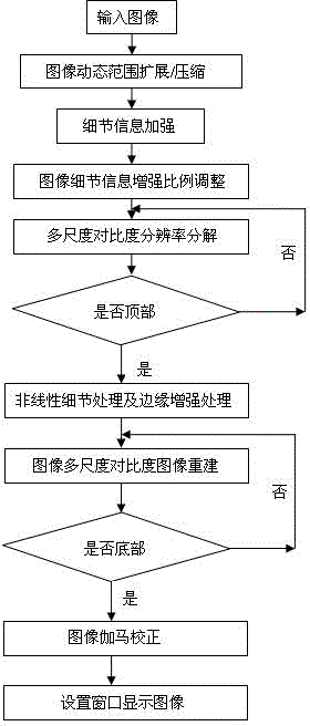
[0022] refer to image 3 The flowchart is characterized in that: using image dynamic range compression technology and detail enhancement processing technology, all image detail information can be displayed under the same window technology, and the doctor's diagnostic level and diagnostic accuracy can be improved. The specific implementation steps are as follows:

[0023] Step 1. Grayscale image compression and expansion technology. In order to make the grayscale distribution of the image more in line with human visual characteristics, logarithmic transformation is used to expand the image information in the low grayscale area and compress the image information in the high grayscale area. Since the human body information mainly Concentrated in the low grayscale area, so the contrast of the image information in the low grayscale area is improved through logarithmic transformation, and the image in the high grayscale area is compressed. The algorithm formula is as follows:

[002...


PUM


Abstract

The invention discloses a digital X-ray image contrast enhancement processing method, and belongs to the technical field of medical instruments. The digital X-ray image contrast enhancement processing method is characterized by comprising the following steps: according to X-ray imaging characteristics, dynamically expanding an interesting human body area image, and dynamically compressing non-human body image information, thereby effectively increasing the contrast of interesting area images; implementing detail enhancement and multi-scale contrast resolution enhancement on images adjusted within a dynamic range, and adjusting the naked eye watching effect of acquired images, thereby completely displaying human body information to a diagnosis doctor. The digital X-ray image contrast enhancement processing method has the advantages that human body image details are conspicuously highlighted, all information of the images can be watched in the same window, lung marking, lung marking tail end renal vessels, bone marking, muscle levels, brain vessels and the like can be all clearly displayed, and the diagnosis skill level can be greatly improved. In addition, on the basis of airspace image processing, the method is high in processing speed and can be applied to a dynamic image processing system.

Description

technical field [0001] The invention belongs to the technical field of medical equipment. Background technique [0002] X-ray images are currently one of the most widely used medical images in clinical practice. Different ray intensities can be obtained, and the change of ray intensity at the receiving end can be received by the detector, and sent to the computer through A / D conversion, which is presented as a change in grayscale. Therefore, X-ray imaging is to convert the three-dimensional structure The projection imaging of the human body in two-dimensional space is the overlapping image of the inner layer of the human body. Due to the dynamic range of the digital X-ray image (2 10 ~2 16 ) is relatively wide, and the human eye’s recognition ability is limited, and the discrimination ability is lower than 100 gray levels. Therefore, how to obtain more image information that the human eye can recognize from the X-ray image, that is, present all the images in one window De...

Claims


Application Information

Patent Timeline
IPC IPC(8): G06T5/00
Inventor 刘金虎苏娟
Owner NANJING PERLOVE RADIAL VIDEO EQUIP
Why Eureka
Try Eureka