

Anti-AKR1B10 protein monoclonal antibody and applications thereof
A monoclonal antibody, 1B10 technology, applied in the field of medicine and biology
- Summary
- Abstract
- Description
- Claims
- Application Information
AI Technical Summary
Problems solved by technology
Method used
Image

Examples
Embodiment 1
[0062] Example 1: Preparation of mouse-derived anti-AKR1B10 protein monoclonal antibody hybridoma cell line
[0063] Use 1.0 mg of purified AKR1B10 full-length protein antigen, add an equal volume of Freund's adjuvant to enhance the specific immune response of the antigen, and use a 5ml syringe to repeatedly pump the above mixture to emulsify it to reach a water-in-oil state. Two healthy males aged 6-8 weeks were selected and immunized three times, 300 μL per mouse. Each time, the emulsified AKR1B10 specific antigen was injected into the subcutaneous and abdominal cavity of the mouse, and the immunization was carried out every 2 weeks. The dose and route of immunization 3 days before cell fusion, the antigen was directly mixed with PBS without adding any adjuvant, and injected directly into the peritoneal cavity of mice for booster immunization. Then, under aseptic conditions, the mouse spleen that had been bled was taken out, ground into a cell suspension, placed in a 50 mL...
Embodiment 2
[0065] Example 2: Preparation of Sandwich Time-Resolved Fluorescence Immunoassay (TRFIA) Kit for Detection of AKR1B10 Protein
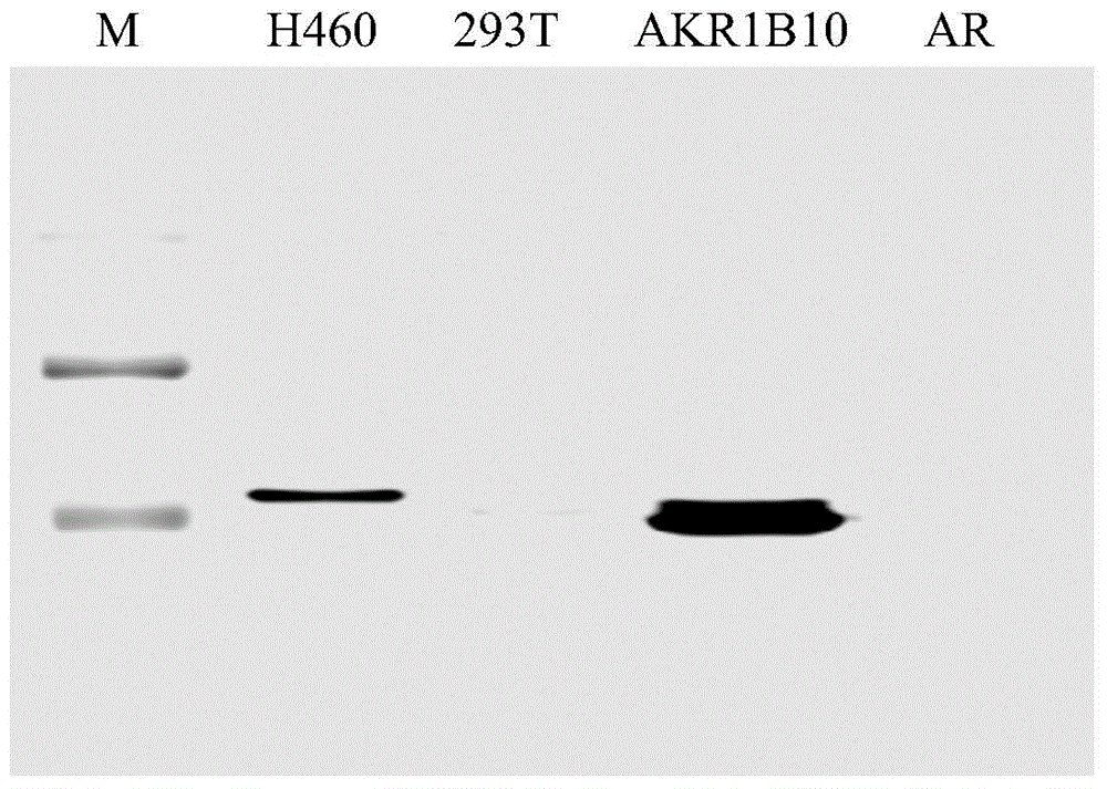
[0066] The monoclonal antibody obtained in Example 1 (that is, the AKR1B10 monoclonal antibody extracted and purified from the preserved 8-DC-1 and 9-DC-2 cell lines) was used as a detection antibody and labeled with biotin; wild-type full-length The AKR1B10 polyclonal antibody obtained after immunizing goats with AKR1B10 protein was used as a capture antibody to construct a sandwich time-resolved fluorescence immunoassay (TRFIA) kit. The sensitivity and specificity of the capture antibody and detection antibody obtained by the present invention are very strong, such as Figure 5 As shown, the time-resolved fluorescence immunoassay (TRFIA) could detect 0.3125 ng / ml of AKR1B10 protein. The biotin labeling kit was purchased from Pierce Company. Streptavidin-Eu was purchased from Perkin Elmer. The time-resolved fluorescent immunoassay (TRFIA) plate ...
Embodiment 3
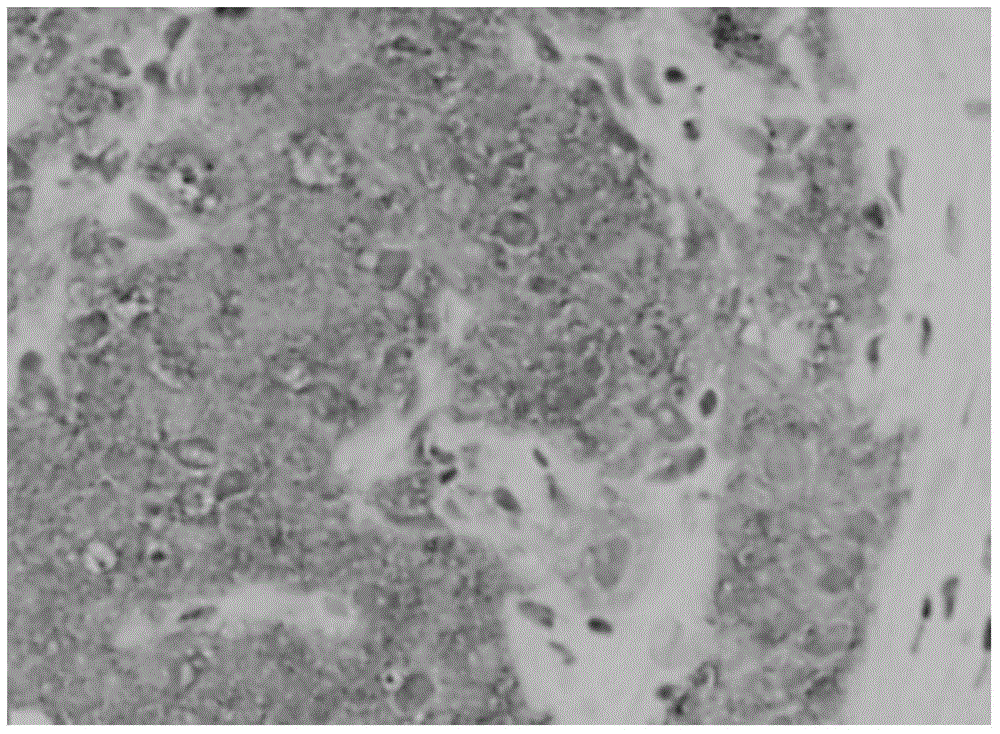
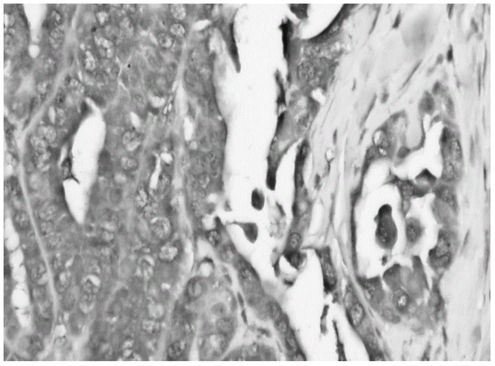
[0068] Embodiment 3: normal crowd and breast cancer patient, liver cancer patient serum AKR1B10 concentration
[0069] Detection of serum samples: Serum samples were diluted 1:10 with PBS, and 100 μl of sample diluent or standard was added to each well (different concentrations of AKR1B10 antigen: 0, 0.3125, 0.625, 1.25, 2.5, 5, 10, 20 ng / ml ), 37oC, 60 minutes; shake off the liquid, wash 5 times with washing buffer, and air dry; dilute the detection antibody with antibody diluent at 1:500, add 100 μl detection antibody diluent to each well, 37oC, 60 minutes; shake Remove liquid, wash 5 times with washing buffer, and air dry; dilute Streptavidin-Eu with antibody diluent at 1:80000, add 100 μl Streptavidin-Eu dilution to each well, 37oC, 60 minutes; shake off liquid, wash with washing buffer 5 times, and air-dried; 100 μl was added to the reaction solution; the OD value was measured with a microplate reader at a wavelength of 450 nm. A standard curve was drawn, and accordi...
PUM
 Abstract
Description
Claims
Application Information