Double-stained kit for auxiliary diagnosis of benign or malignant hepatocellular tumor and application thereof

A malignant tumor, auxiliary diagnosis technology, applied in the direction of measuring devices, instruments, scientific instruments, etc., can solve the problems of lack of specificity and sensitivity, little help in diagnosis, difficulty, etc., to increase the readability and accuracy. Effect

Inactive Publication Date: 2015-06-24
GUANGZHOU LBP MEDICINE SCI & TECH
View PDF4 Cites 13 Cited by
  • Summary
  • Abstract
  • Description
  • Claims
  • Application Information

AI Technical Summary

Problems solved by technology

It is difficult to distinguish HCC from other liver masses solely by morphology
IHC can play an important role in the auxiliary diagnosis of benign and malignant liver masses, but commonly used antibodies such as CEA (polyclonal), CD10, and AFP lack specificity and sensitivity, and are of little help to the diagnosis

Method used


Image

  • Double-stained kit for auxiliary diagnosis of benign or malignant hepatocellular tumor and application thereof
  • Double-stained kit for auxiliary diagnosis of benign or malignant hepatocellular tumor and application thereof
  • Double-stained kit for auxiliary diagnosis of benign or malignant hepatocellular tumor and application thereof

Examples


Embodiment 1

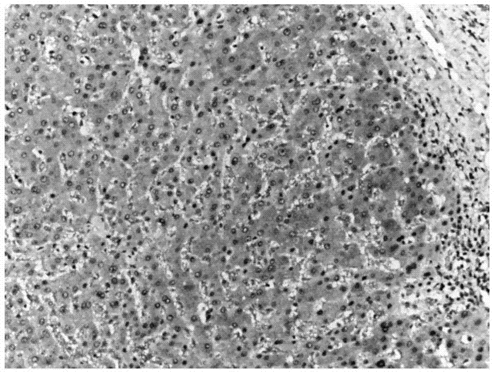
[0046] The research objects were formalin-fixed-paraffin-embedded hepatic nodular hyperplastic tissues (provided by Guangzhou Da'an Clinical Laboratory Center Co., Ltd.). The steps of immunohistochemical experiment are as follows:

[0047] (1) Dewaxing and hydration: Before dewaxing, place the tissue slices on a 60°C oven for 2 hours. Soak in xylene for 10 minutes twice, in absolute ethanol for 5 minutes twice, in 95% ethanol for 5 minutes once, and in distilled water for 1 minute once.

[0048] (2) Antigen retrieval: Boil the tissue section in step (1) in EDTA (pH 9.0) antigen retrieval solution, cool, rinse with tap water, and soak in distilled water.

[0049] (3) Sealing: incubate the tissue section in step (2) with 3% hydrogen peroxide at room temperature for 10 minutes, rinse with distilled water, wash and soak in TBS buffer.

[0050] (4) Double staining: In step (3), the tissue section is dripped with the first antibody, that is, Arginase-1 and Glypican-3 mixed primary...

Embodiment 2

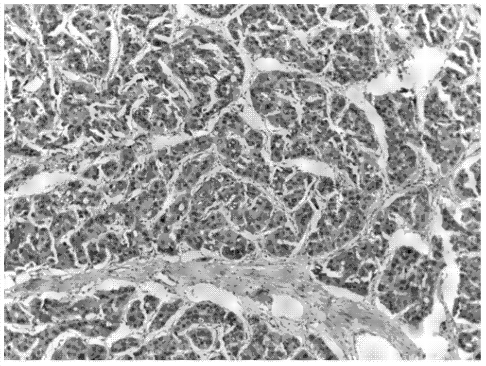
[0058] The research object was formalin-fixed-paraffin-embedded poorly differentiated hepatocellular carcinoma (provided by Guangzhou Da'an Clinical Laboratory Center Co., Ltd.). The steps of immunohistochemical experiment are as follows:

[0059] (1) Dewaxing and hydration: Before dewaxing, place the tissue slices on a 60°C oven for 2 hours. Soak in xylene for 10 minutes twice, in absolute ethanol for 5 minutes twice, in 95% ethanol for 5 minutes once, and in distilled water for 1 minute once.

[0060] (2) Antigen retrieval: the tissue section in step (1) was boiled in EDTA (pH 9.0) antigen retrieval solution, cooled, rinsed with tap water, and soaked in distilled water.

[0061] (3) Sealing: incubate the tissue section in step (2) with 3% hydrogen peroxide at room temperature for 10 minutes, rinse with distilled water, wash and soak in TBS buffer.

[0062] (4) Double staining: In step (3), the tissue section is dripped with the first antibody, that is, Arginase-1 and Glypi...

Embodiment 3

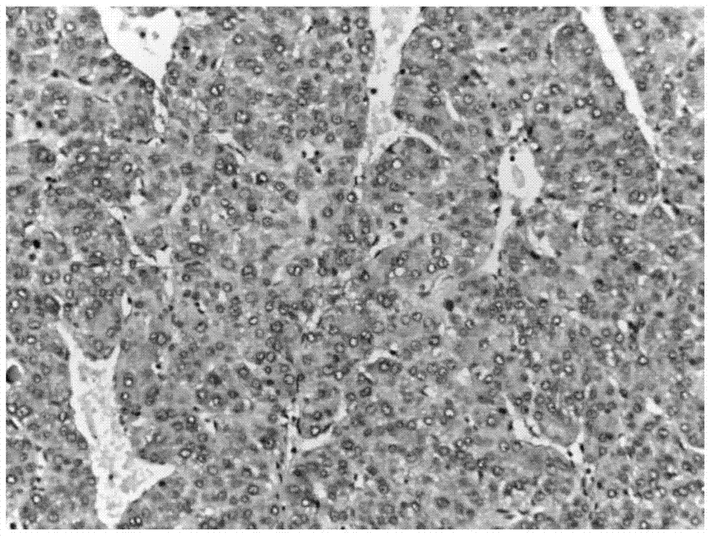
[0070] The research object was formalin-fixed-paraffin-embedded well-differentiated hepatocellular carcinoma (provided by Guangzhou Da'an Clinical Laboratory Center Co., Ltd.). The steps of immunohistochemical experiment are as follows:

[0071] (1) Dewaxing and hydration: Before dewaxing, place the tissue slices on a 60°C oven for 2 hours. Soak in xylene for 10 minutes twice, in absolute ethanol for 5 minutes twice, in 95% ethanol for 5 minutes once, and in distilled water for 1 minute once.

[0072] (2) Antigen retrieval: Boil the tissue section in step (1) in EDTA (pH 9.0) antigen retrieval solution, cool, rinse with tap water, and soak in distilled water.

[0073] (3) Sealing: incubate the tissue section in step (2) with 3% hydrogen peroxide at room temperature for 10 minutes, rinse with distilled water, wash and soak in TBS.

[0074] (4) Double staining: In step (3), the tissue section is dripped with the first antibody, that is, Arginase-1 and Glypican-3 mixed primary ...


PUM

PropertyMeasurementUnit
thicknessaaaaaaaaaa

Abstract

The invention discloses a double-stained kit for auxiliary diagnosis of benign or malignant hepatocellular tumor and application thereof. The kit comprises the following components: mixed primary antibody of Arginase-1 and Glypian-3, mixed second antibody of alkaline phosphatase conjugated goat anti-rabbit second antibody and horse radish peroxidase labeled goat anti -mouse second antibody, chromogenic reagent AP-Red and DAB (Diaminobenzidine), and TBS (Tris buffer saline) buffer solution. Two immunohistochemical labels provided by the invention are used for auxiliary diagnosis of benign or malignant hepatocellular tumor and differentiation degrees of tumor, the two labels are displayed through staining label by different detection systems and utilizing the differences of target cells stained by each label, information quantity and differential staining on the same radiograph are improved, readability and accuracy of radiograph reading are increased, so that the double-stained kit has important significance to identification of benign or malignant hepatocellular tumor and differentiation degrees of tumor.

Description

technical field [0001] The present invention relates to the field of immunohistochemistry, more specifically, to a double-staining kit for auxiliary diagnosis of benign and malignant hepatic tumors and its application and immunohistochemical double-staining for auxiliary differential diagnosis of benign and malignant hepatic tumors Detection method. Background technique [0002] Liver cancer is a malignant tumor that seriously endangers human life and health. Its prevalence rate is increasing year by year in my country. The surgical resection rate of the disease is low, and the 5-year survival rate is less than 5%. It is now the second leading cause of cancer death in my country. . The etiology and clinical manifestations of primary liver cancer are relatively complex and lack specificity. Distant metastasis often occurs at the time of clinical diagnosis, which leads to delay in treatment and low postoperative survival rate. Therefore, the prevention, diagnosis and treatmen...

Claims


Application Information

Patent Timeline
Patent Type & Authority Applications(China)
IPC IPC(8): G01N33/574
CPCG01N33/57438
Inventor 杨鉴李玉闫有臣张海明
Owner GUANGZHOU LBP MEDICINE SCI & TECH
Why Eureka
Try Eureka