
A method for registration of 3D CT and ultrasound liver images based on fast convex optimization algorithm

A convex optimization algorithm and CT image technology, applied in image enhancement, image analysis, image data processing, etc., can solve problems such as large image differences, inability to achieve high precision, automatic and accurate real-time registration difficulties, etc.

Active Publication Date: 2018-05-29
ZHEJIANG DE IMAGE SOLUTIONS CO LTD
View PDF2 Cites 0 Cited by
  • Summary
  • Abstract
  • Description
  • Claims
  • Application Information

AI Technical Summary

Problems solved by technology

[0003] The registration of ultrasound images and CT images is still an international problem, and automatic and accurate real-time registration is still difficult in actual clinical applications
There are many difficulties in liver ultrasound-CT registration, mainly including the following aspects: (1) Because the images of CT and ultrasound are very different, and the structures that can be visualized in CT may not be clearly seen in ultrasound, It is difficult to set the similarity measure of the two modalities, which belongs to the multimodal registration problem in the registration problem
(2) Due to the narrow field of view of ultrasound, the liver is the largest organ in the abdomen, and because of the occlusion of the abdominal bone, only part of the liver can be displayed in ultrasound images, and the complete liver cannot be displayed like CT; (3) Respiratory movement Effect on Image Registration Accuracy
Although this kind of registration by external markers is fast, it cannot achieve high accuracy, especially when dealing with local deformation.
These methods have their own advantages and disadvantages, and cannot completely solve the above problems.

Method used


Image

  • A method for registration of 3D CT and ultrasound liver images based on fast convex optimization algorithm
  • A method for registration of 3D CT and ultrasound liver images based on fast convex optimization algorithm
  • A method for registration of 3D CT and ultrasound liver images based on fast convex optimization algorithm

Examples


Embodiment Construction

[0043] Below in conjunction with accompanying drawing and specific embodiment the present invention is described in further detail:

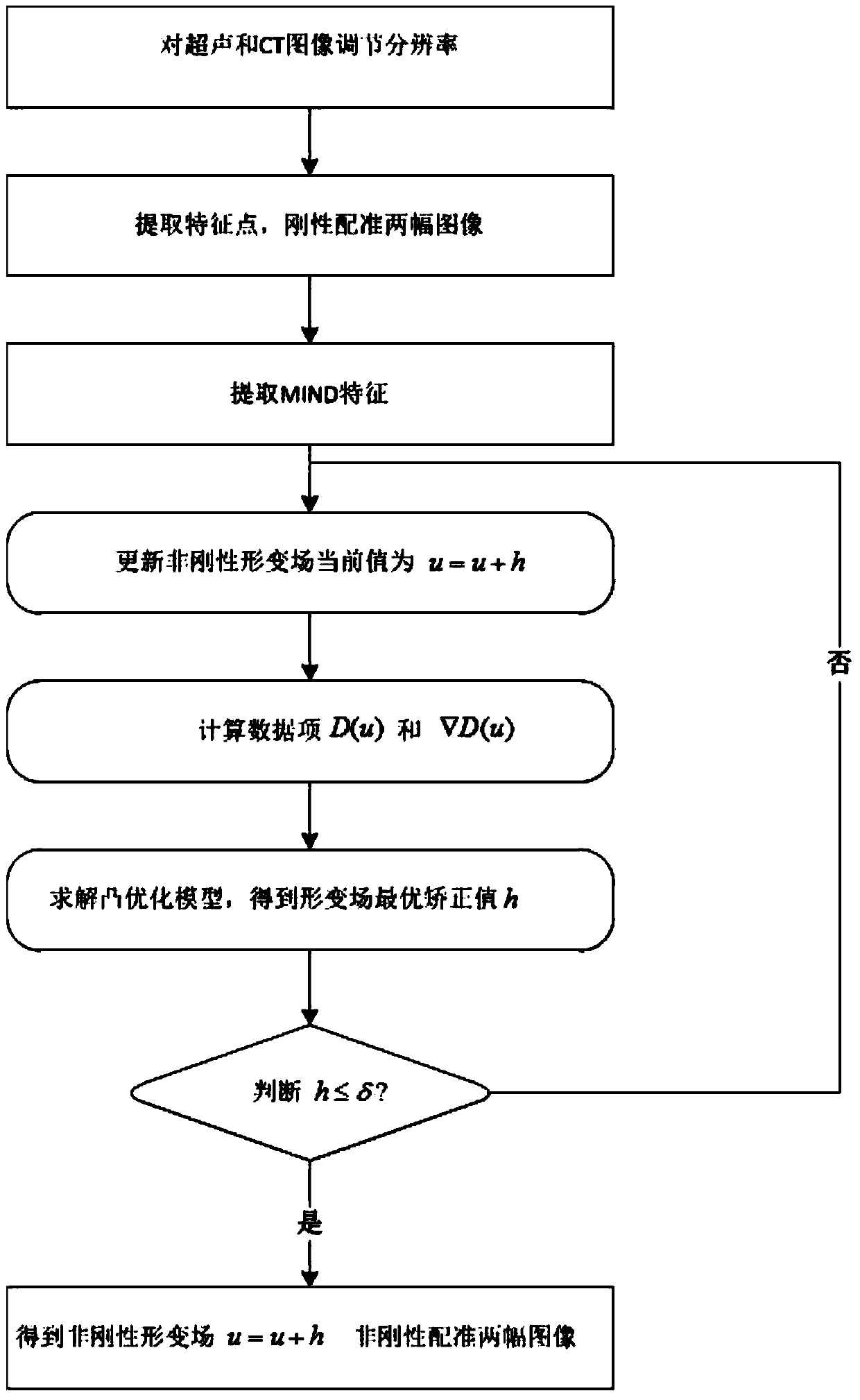
[0044] figure 1 It is a flow chart of registering 3D CT and ultrasound liver images based on the fast convex optimization algorithm, specifically including the following process:

[0045] Step A: For the input liver CT image I C (x) and ultrasound image I U (x) Adjust the window width and level so that the image display value is within [0, 255]. I C (x) has a size of 512×512×58, and the size of each voxel is 0.79mm×0.79mm×3mm, and the I C (x) size becomes 1094×1094×115, each voxel size is 0.37mm×0.37mm×1.5mm. I U (x) has a size of 300×300×1275, and the size of each voxel is 0.37mm×0.37mm×0.10mm. By downsampling, I U The size of (x) becomes, 300×300×85, and the size of each voxel is 0.37mm×0.37mm×1.5mm. The symbols are all the same as those defined in the step narration part of the description.

[0046] Step B: For the rough registration...


PUM


Abstract

The invention relates to the field of medical image post-processing and aims to provide a method for registering three-dimensional CT and ultrasound liver images based on a fast convex optimization algorithm. The method of registering three-dimensional CT and ultrasound liver images based on a fast convex optimization algorithm includes the following processes: adjusting the resolution of ultrasound and CT images to the same; coarse registration based on rigid body transformation of ultrasound and CT images; extracting multi-modality Unified feature information for image registration; calculate the gradient field of D(u) and D(u) in the data item under the current non-rigid deformation field u(x), solve the model for each step of the stepwise convex optimization method, and obtain the deformation With the optimal field correction value h(x), the deformation field is updated until h(x) is very small; according to the solved non-rigid deformation field, the ultrasound image is transformed and registered with the CT image. By establishing a reasonable model, the present invention designs a fast and accurate three-dimensional ultrasound-CT liver image registration algorithm to improve the accuracy, safety and effectiveness of ablation surgery.

Description

technical field [0001] The invention relates to the field of post-processing of medical images, in particular to a method for registering three-dimensional CT and ultrasonic liver images based on a fast convex optimization algorithm. Background technique [0002] At present, liver disease is a disease with a relatively high incidence rate clinically, which directly threatens people's lives. In the treatment of liver cancer, local ablation therapy represented by radiofrequency ablation has become the third major liver cancer treatment method besides surgical resection and interventional therapy. Due to the advantages and disadvantages of ultrasound imaging, ultrasound-CT fusion is often used in ablation procedures to guide surgical needle insertion. The key technology is the registration of CT-ultrasound multimodal images. [0003] The registration of ultrasound images and CT images is still an international problem, and automatic and accurate real-time registration is stil...

Claims


Application Information

Patent Timeline
Patent Type & Authority Patents(China)
IPC IPC(8): G06T3/00G06T7/32
CPCG06T7/30G06T7/344G06T2207/10132G06T2207/10081G06T2207/30056G06T3/14
Inventor 孔德兴袁景胡佩君
Owner ZHEJIANG DE IMAGE SOLUTIONS CO LTD
Learn More