Method for sorting hepatic vessels

A technology of liver blood vessels and blood vessels, which is applied in the field of classification of liver blood vessels, can solve the problems of non-vascular tissue mixing, spending more energy, and losing details of blood vessels, so as to improve accuracy, improve the actual medical reference application value, and facilitate preoperative Estimated effect

Inactive Publication Date: 2015-12-09
QINGDAO HISENSE MEDICAL EQUIP
View PDF5 Cites 18 Cited by
  • Summary
  • Abstract
  • Description
  • Claims
  • Application Information

AI Technical Summary

Problems solved by technology

However, due to factors such as medical imaging itself and individual patient liver tumor location and blood vessel variation, the actual CT image quality is not high. Three-dimensional vascular images often show the overlapping of hepatic artery, portal vein and hepatic vein, as well as other non-vascular tissues. Usually, medical staff can only determine the roots of the hepatic artery, hepatic vein and portal vein according to the different anatomical structures , and then classify the hepatic artery, hepatic vein, and portal vein according to the thickness, connectivity, and shape of the blood vessels, which requires more energy for medical staff, and the sampling method above will often lose many details of blood vessels

Method used


Image

  • Method for sorting hepatic vessels
  • Method for sorting hepatic vessels
  • Method for sorting hepatic vessels

Examples


Embodiment 1

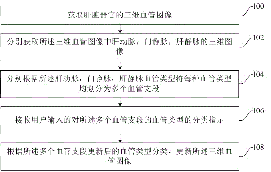
[0023] An embodiment of the present invention provides a method for classifying liver blood vessels, referring to figure 1 , the method includes the following steps:

[0024] Step 100, acquiring a three-dimensional blood vessel image of the liver organ.

[0025] Wherein, the three-dimensional blood vessel image is obtained by modeling according to a group of two-dimensional CT images in the same period. For example, as described in the background art, an arterial vessel model can be created by using an image in the arterial phase, a portal vein model can be created by using an image in the portal venous phase, and a hepatic vein model can be created by using an image in the equilibrium phase.

[0026] Specifically, for the three-dimensional blood vessel image of the liver organ acquired in step 100, it specifically includes as follows figure 2 as shown,

[0027] 100a. Perform blood vessel segmentation on each two-dimensional blood vessel image in a group of two-dimensional...

Embodiment 2

[0053] Embodiment 2 of the present invention is an improvement on the method for classifying liver blood vessels on the basis of Embodiment 1.

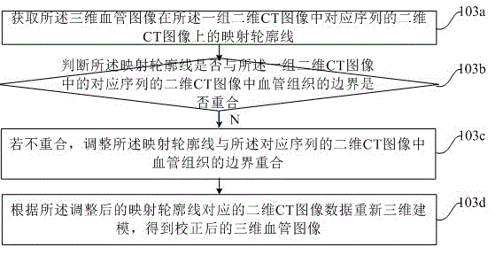
[0054] Specifically, before step 104, dividing each blood vessel type into a plurality of blood vessel branches according to the hepatic artery, portal vein, and hepatic vein respectively, step 103, performing two-dimensional image processing on the three-dimensional blood vessel image Map correction.

[0055] Since in step 100a of the first embodiment, when performing vessel segmentation on each two-dimensional vessel image according to the region growing algorithm, sometimes similar pixels that do not belong to the vessel region are also classified into the vessel region, therefore, the three-dimensional vessel image is divided into each Map the two-dimensional blood vessel image, adjust the inaccurate position, so that the reconstructed three-dimensional blood vessel image is more accurate, so as to conform to the actual vascular s...

Embodiment 3

[0080] The embodiment of the present invention is an improvement on the basis of the first embodiment or the combination of the first embodiment and the second embodiment.

[0081] Specifically, before step 104 of the first embodiment, dividing each blood vessel type into a plurality of blood vessel branches according to the hepatic artery, portal vein, and hepatic vein respectively, or in step 103 of the second embodiment, dividing the three-dimensional After the two-dimensional image mapping correction is performed on the blood vessel image, another step 105 of correcting the three-dimensional blood vessel image is included, removing non-vascular tissue in the three-dimensional blood vessel image.

[0082] Since the embodiment of the present invention is mainly aimed at the three-dimensional image of the blood vessel, the non-vascular tissue in the three-dimensional blood vessel image may be removed first, so as to obtain a more accurate three-dimensional blood vessel image. ...


PUM


Abstract

The invention provides a method for sorting hepatic vessels. The method for sorting hepatic vessels comprises redistricting a plurality of branches of a vessel according to the types of hepatic vessels; receiving the sorting indication of a user for the vessel types of the plurality of branches of a vessel; and updating three dimensional vessel images according to the sorting of updated vessel types of the plurality of branches of a vessel. The method for sorting hepatic vessels can improve the accuracy for sorting vessels in the three dimensional vessel images of liver organs so as to improve the practical medical reference application value of the three dimensional vessel images. The method for sorting hepatic vessels can be used for three dimensional modeling image processing in the medical field.

Description

technical field [0001] The invention relates to the technical field of medical image processing, in particular to a method for classifying liver blood vessels. Background technique [0002] With the continuous improvement of medical technology, in order to obtain the location of the patient's lesion more accurately, conventional abdominal CT (full name in English: Computed Tomography Chinese: computerized tomography) enhanced three-phase dynamic scanning of the human body is used in the prior art to obtain arterial Three-phase images of phase, portal vein phase, and equilibrium phase, and then use three-dimensional reconstruction technology to image the hepatic artery, portal vein, and hepatic vein angiography, and analyze the distribution structure and variation of the three in the liver. For liver segmentation, liver tumor resection has important guiding significance. [0003] If the quality of the CT images provided is good, the arterial phase images can be used to creat...

Claims


Application Information

Patent Timeline
Patent Type & Authority Applications(China)
IPC IPC(8): G06K9/62
CPCG06F18/2431G06F18/00
Inventor 刘丽丽陈永健
Owner QINGDAO HISENSE MEDICAL EQUIP
Why Eureka
Try Eureka