

Kit for detecting C-reactive protein through fluorescent immunochromatography
A technology of fluorescence immunochromatography and detection kits, applied in biological testing, measuring devices, analytical materials, etc., can solve the problems of narrow linearity, low sensitivity, unfavorable for high-throughput automatic detection, etc., and achieve the effect of reducing errors
- Summary
- Abstract
- Description
- Claims
- Application Information
AI Technical Summary
Problems solved by technology
Method used
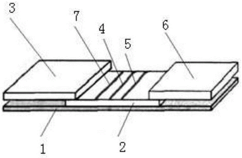
Image

Examples
Embodiment 1
[0046] AlexaFluor660 from Lifetechnologies was used for fluorescein, which was dissolved in DMSO and then adjusted to a concentration of 10 mg / ml as a fluorescein storage solution. The fluorescein was activated by EDC / NHS cross-linking method (the molar ratio of fluorescein to the total amount of chemical cross-linking agent was 1:0.14). The fluorescein storage solution was mixed with EDC and NHS, and 50 mM pH6.0 MES buffer was used as the reaction medium, and incubated at 37° C. for 60 minutes to obtain activated fluorescein.
[0047] Take 1 mg of activated fluorescein and add 20 mg of mouse anti-human CRP monoclonal antibody I (T1) to incubate at 37°C for 60 minutes. After that, free fluorescein was dialyzed with 50 mM Tris-HCl buffer pH7.4, and the dialysate was changed every 6 hours for a total of three times. Collect the liquid in the dialysis bag, use Tris-HCl 50mM / L, 1% BSA, 0.6% Triton X-100, 0.9% NaCl, 0.05% NaN after ultrafiltration and concentration 3 Fluorescein ...
Embodiment 2
[0057] AlexaFluor 660 from Lifetechnologies was used for fluorescein, which was dissolved in DMSO and then adjusted to a concentration of 5 mg / ml as a fluorescein storage solution. The fluorescein was activated by EDC / NHS cross-linking method (the molar ratio of fluorescein to the total amount of chemical cross-linking agent was 1:0.14). The luciferin storage solution was mixed with EDC and NHS, and the activated luciferin was obtained by incubating at 36° C. for 10 minutes with 50 mM pH6.2 MES buffer as the reaction medium.
[0058] Take 1 mg of activated fluorescein and add 5 mg of mouse anti-human CRP monoclonal antibody I (T1) to incubate at 36° C. for 10 minutes. After that, free fluorescein was dialyzed with 50 mM Tris-HCl buffer pH7.4, and the dialysate was changed every 6 hours for a total of three times. Collect the liquid in the dialysis bag, use Tris-HCl 5mM / L, 2.3% BSA, 0.1% Triton X-100, 0.9% NaCl, 0.43% NaN after ultrafiltration and concentration 3 Fluorescein ...
Embodiment 3
[0066] The fluorescein was AlexaFluor 660 from Lifetechnologies, which was dissolved in DMSO and then adjusted to a concentration of 7 mg / ml as a fluorescein storage solution. The fluorescein was activated by EDC / NHS cross-linking method (the molar ratio of fluorescein to the total amount of chemical cross-linking agent was 1:0.14). The luciferin stock solution was mixed with EDC and NHS, and 50 mM pH6.4 MES buffer was used as the reaction medium, and incubated at 38° C. for 120 minutes to obtain activated luciferin.
[0067] Take 1 mg of activated fluorescein and add 50 mg of mouse anti-human CRP monoclonal antibody I (T1) to incubate at 38° C. for 120 minutes. After that, free fluorescein was dialyzed with 50 mM Tris-HCl buffer pH7.4, and the dialysate was changed every 6 hours for a total of three times. Collect the liquid in the dialysis bag, use Tris-HCl 200mM / L, 1.7% BSA, 2% Triton X-100, 0.9% NaCl, 0.85% NaN after ultrafiltration and concentration 3 Fluorescein Antibo...
PUM
 Abstract
Description
Claims
Application Information