

PET/CT macroscopical digital information and pathological microscopic information matching method
A technology of digital information and information matching, applied in image data processing, instrumentation, computing, etc., can solve the problems of limiting the development of PET/CT and the inability to detect endogenous tumor markers by immunohistochemistry
- Summary
- Abstract
- Description
- Claims
- Application Information
AI Technical Summary
Problems solved by technology
Method used
Image

Examples
Embodiment 1
[0029] A method for matching PET / CT macroscopic digital information with pathological microscopic information, comprising the following steps:
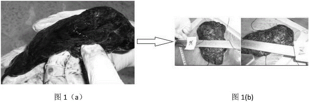
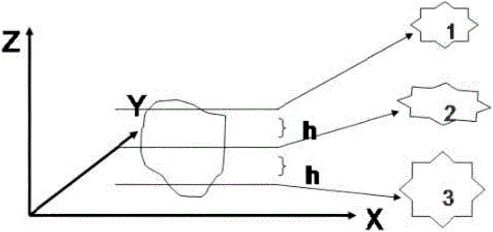
[0030] Step 1: As shown in Figure 1(a) and Figure 1(b), restore the postoperative specimen to the in vivo state, and then immerse it in formalin for 24 hours; The resected lung tissue is marked with X, Y, and Z in the natural state of the body, and the lung tissue is isolated to form a specimen, and the specimen is positioned according to the three-dimensional direction in the body, and the specimen is restored to the in vivo state;
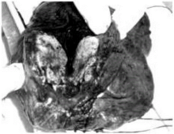
[0031] Step 2: If Figure 2(a)-Figure 2(c) As shown, review the preoperative PET / CT image, and the cross-section is formed between the highest level of tracer uptake in the PET / CT image and the carina (in this embodiment, it is the lesion in the lung, so the carina is selected; if it is For intracerebral lesion scanning, select the blood vessel indicated by the specimen), cut the specimen from the middle...
Embodiment 2
[0039] A method for matching PET / CT macroscopic digital information with pathological microscopic information, comprising the following steps:
[0040] Step 1: As shown in Figure 1(a) and Figure 1(b), restore the postoperative specimen to the in vivo state, and then immerse it in formalin for 16 hours; The resected lung tissue is marked with X, Y, and Z in the natural state of the body, and the lung tissue is isolated to form a specimen, and the specimen is positioned according to the three-dimensional direction in the body, and the specimen is restored to the in vivo state;
[0041] Step 2: If Figure 2(a)-Figure 2(c) As shown, review the preoperative PET / CT image, and the cross-section is formed between the highest level of tracer uptake in the PET / CT image and the carina (in this embodiment, it is the lesion in the lung, so the carina is selected; if it is For intracerebral lesion scanning, select the blood vessel indicated by the specimen), cut the specimen from the middle...
PUM
 Abstract
Description
Claims
Application Information