Microflow device based on SERS (surface enhanced Raman scattering) technology to detect specific antigens in serum

A microfluidic device and technical detection technology, which is applied in the fields of spectroscopy and biological analysis, can solve the problems of difficult quantitative testing, large display contrast, and high price, and achieve the advantages of easy testing and portability, less testing samples, and less time-consuming testing. Effect

Inactive Publication Date: 2015-12-30
CHINA JILIANG UNIV
View PDF5 Cites 5 Cited by
  • Summary
  • Abstract
  • Description
  • Claims
  • Application Information

AI Technical Summary

Problems solved by technology

Immunofluorescence microscopy is expensive, it is difficult to use for quantitative testing, and the technical procedures are complicated
It is difficult to find a suitable enzyme-catalyzed substrate for enzyme-linked immunoassay, which makes it difficult to apply, and there is a large error in display comparison
Polymerase chain reaction detection technology is difficult, the requirements for equipment are too high, the operation is cumbersome, and the price is expensive
These shortcomings limit the application of these methods and can only be used for laboratory testing

Method used


Image

  • Microflow device based on SERS (surface enhanced Raman scattering) technology to detect specific antigens in serum
  • Microflow device based on SERS (surface enhanced Raman scattering) technology to detect specific antigens in serum
  • Microflow device based on SERS (surface enhanced Raman scattering) technology to detect specific antigens in serum

Examples


Embodiment 1

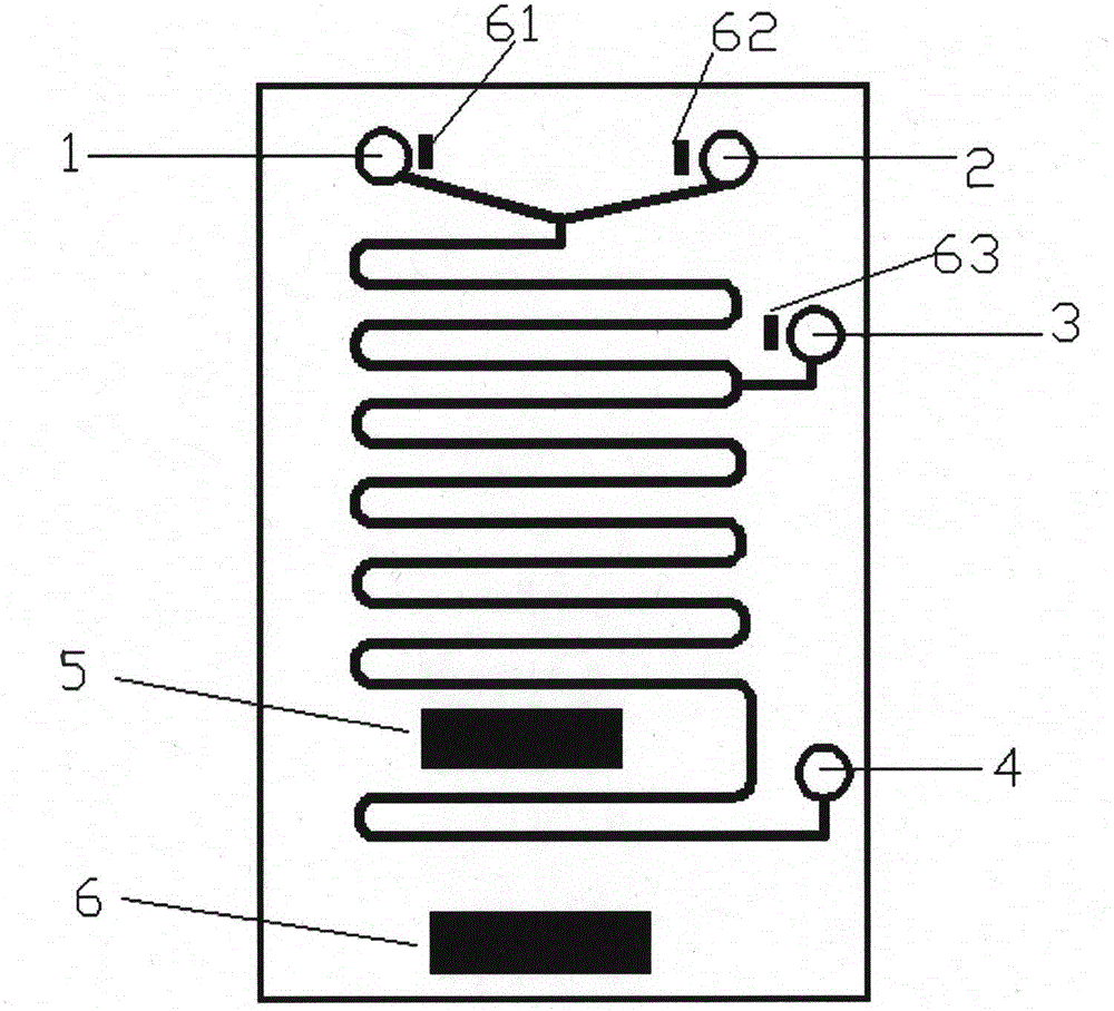
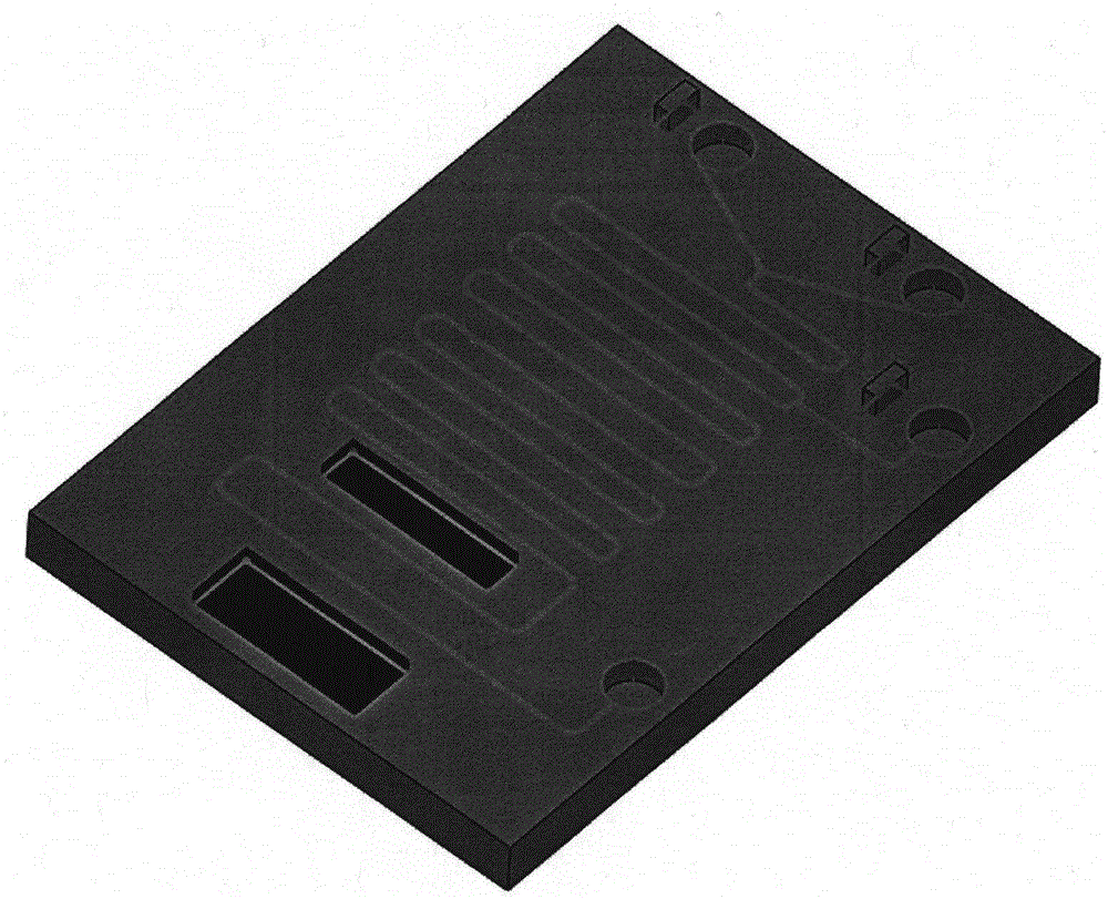
[0033] 1) Preparation of microfluidic device: use quartz glass as the bottom layer, apply a layer of photoresist on it, and use soft photolithography to carve the shape of the microfluidic channel on the photoresist, using polydimethylsiloxane (PDMS) Process to generate a microfluidic channel layer, heating and curing the bottom layer and the middle layer, all channels are 200-250 μm wide and 80-100 μm high, and the microfluidic channel layer and the corresponding uppermost layer are encapsulated by hot pressing, and the microfluidic device packaging layer The SERS detection unit and sensor controller are placed on the upper patch. The bottom layer of the microfluidic device is 3mm, the thickness of the packaging layer is 3mm, and the size of the whole microfluidic device is 6cm×8cm×0.8mm.

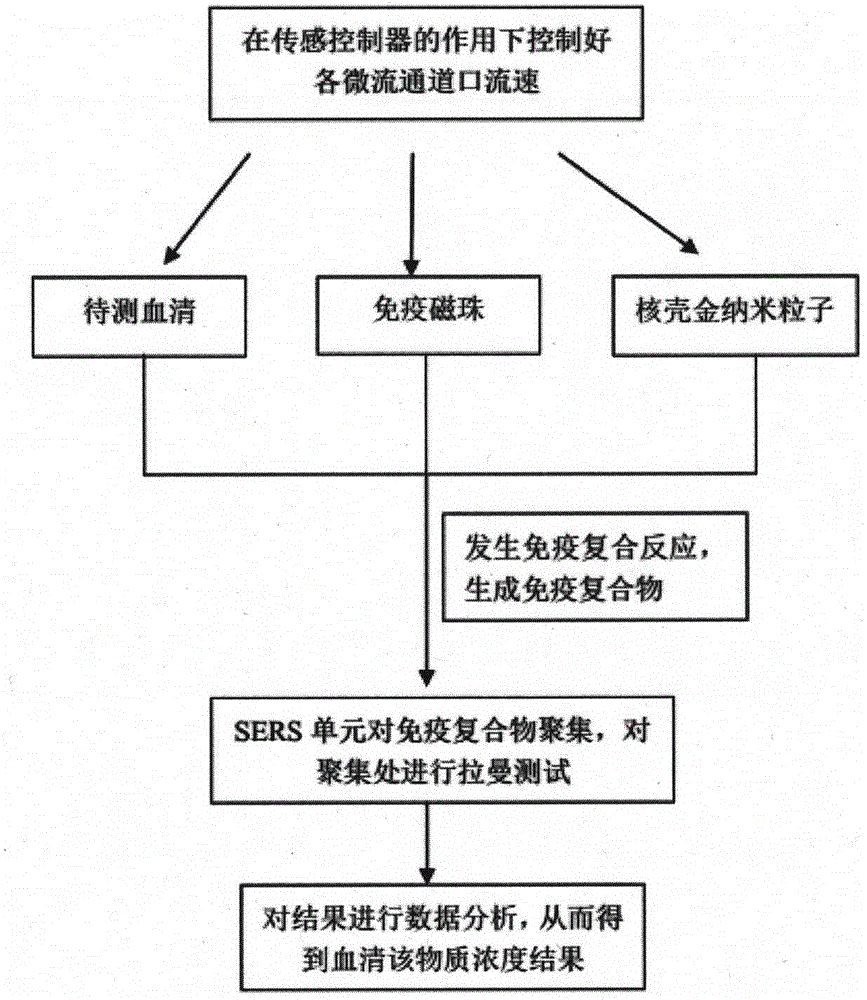
[0034] 2) Preparation of core-shell nanoparticles combined with the serum antigen to be tested: gold nanoparticles with a particle size of about 50 nm were obtained by the chloroaurate and...

Embodiment 2

[0039] Taking the detection of anthrax virus in serum as an example, Bacillus anthracis is mainly composed of γ-polyglutamic acid (PGA), and the content of Bacillus anthracis in the human body can be determined by detecting the content of PGA in serum.

[0040] 1) Preparation of microfluidic device: use quartz glass as the bottom layer, apply a layer of photoresist on it, and use soft photolithography to carve the shape of the microfluidic channel on the photoresist, using polydimethylsiloxane (PDMS) Process to generate a microfluidic channel layer, heating and curing the bottom layer and the middle layer, all channels are 200-250 μm wide and 80-100 μm high, and the microfluidic channel layer and the corresponding uppermost layer are encapsulated by hot pressing, and the microfluidic device packaging layer The SERS detection unit and sensor controller are placed on the upper patch. The bottom layer of the microfluidic device is 3mm, the thickness of the encapsulation layer is ...


PUM

PropertyMeasurementUnit
particle diameteraaaaaaaaaa
thicknessaaaaaaaaaa
diameteraaaaaaaaaa

Abstract

Disclosed is a microflow device based on SERS (surface enhanced Raman scattering) technology to detect specific antigens in serum. The microflow device is of a sandwich structure and is mainly composed of three microflow liquid inlets, one microflow liquid outlet, an SERS detection unit and a sensing controller. Under control of a control sensor, microflow liquid at each microflow passage inlet is in antigen antibody reaction in a passage, the SERS detection unit enables an immune complex in generated microflow liquid to be separated and gathered in a microflow passage nearest to the SERS detection unit, a Raman test is performed on an immune complex aggregate, and concentration of a to-be-detected antigen in serum can be acquired by processing and analyzing acquired SERS signal strength data to judge whether a disease exists or not. The microflow device has the advantages of low time consumption, small sample loss, short detection time and reusability.

Description

technical field [0001] The invention belongs to the field of spectroscopy and biological analysis, and in particular relates to the design and application of a microfluidic device for detecting specific antigens in serum based on surface-enhanced Raman scattering technology. Background technique [0002] With environmental problems becoming more and more serious, we are in an environment that is highly susceptible to virus infection. Viruses such as SARS, influenza, and MERS make everyone feel at risk. Most viruses exist in blood. How to detect the content of a certain virus in serum plays an important role in early diagnosis and post-exposure treatment and diagnosis. Known detection methods using antigen-antibody reaction to measure serum virus cells include immunofluorescence microscopy, enzyme-linked immunoassay and polymerase chain reaction detection, but there are certain defects. Immunofluorescence microscopy is expensive, and it is difficult to use in quantitative te...

Claims


Application Information

Patent Timeline
Patent Type & Authority Applications(China)
IPC IPC(8): G01N21/65B01L3/00
Inventor 董前民王鹏辉金尚忠赵春柳梁培陈华才
Owner CHINA JILIANG UNIV
Why Eureka
Try Eureka