Image detection method for rotting oranges caused by penicillium infection
An image detection and indexing image technology, applied in the field of spectral detection, can solve the problems of ignoring feature expression, not suitable for automatic detection of Penicillium digitatum infected fruit, etc., to reduce postharvest losses, simple and effective image processing algorithm, and increase farmers' income Effect
- Summary
- Abstract
- Description
- Claims
- Application Information
AI Technical Summary
Problems solved by technology
Method used
Image
Examples
Embodiment 1
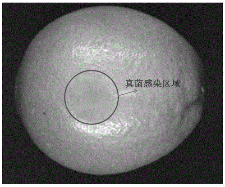
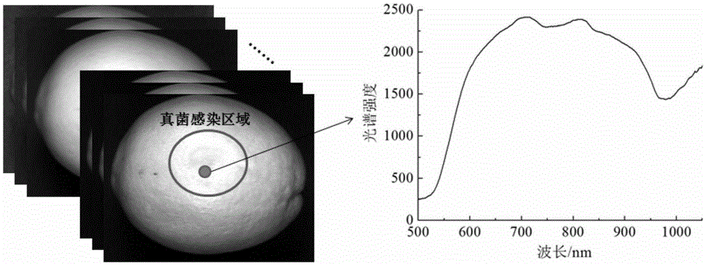
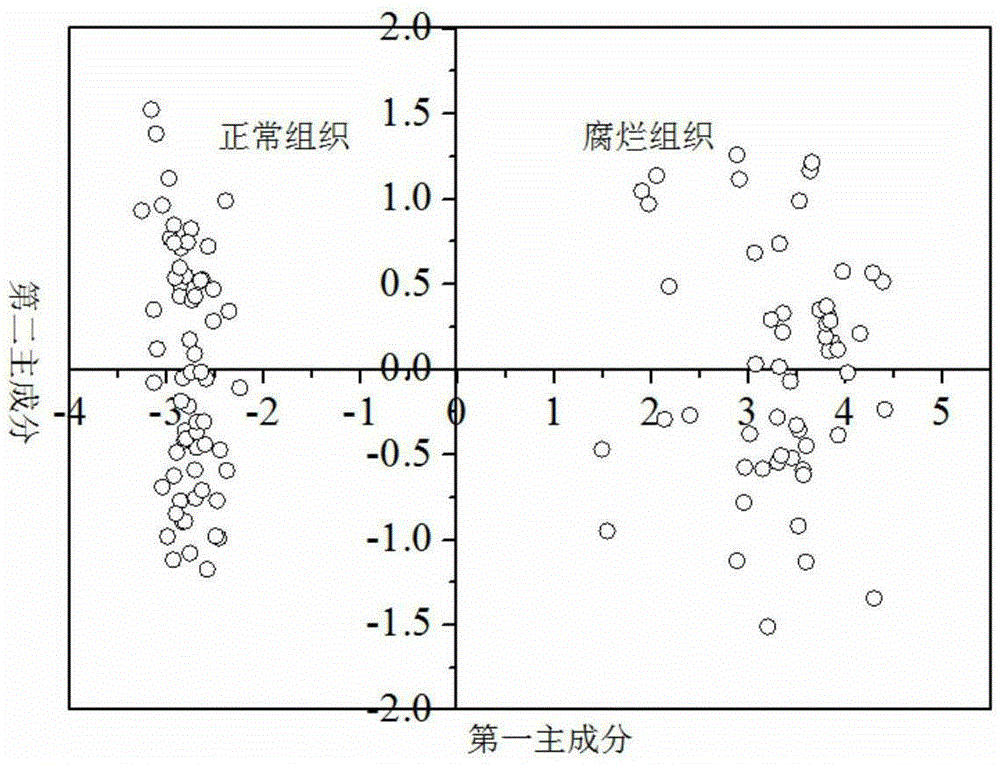
[0057] The invention provides an image detection method of rotten citrus caused by Penicillium infection, which can visualize and image the infected area that is difficult to detect and provide an effective image processing and analysis method. Principal component clustering analysis was performed on the component spectra of the region and the component spectra of fungal infection regions of interest. Prepare 120 citrus fruit oranges. The 120 samples include 60 samples of normal fruit and 60 samples of fungal-infected fruit. The fungal-infected fruit is obtained by artificial inoculation. For the citrus peel tissue of Penicilliumdigitatum, disinfect with 75% medical alcohol for 1min, rinse the spores with sterile water, dissolve the spores in sterile water to form a spore solution, and then use a syringe to inoculate the spore solution into normal fruits, the inoculation depth is about 10mm under the citrus peel . Then place it in a plastic incubator (ambient temperature 25-2...
Embodiment 2
[0060] Based on the characteristic wavelength that embodiment 1 obtains, the method of the present invention comprises the following steps:
[0061] A: Use the visible-near-infrared hyperspectral imaging system (ImSpectorV10E, Spectral Imaging Ltd, Oulu, Finland) to acquire single-wavelength spectral images at four characteristic wavelengths of the citrus fruit to be tested. The four characteristic wavelengths are 575nm, 698nm, 810nm and 969nm ,Such as Figure 5 As shown, it can be seen from the figure that on the one hand, the brightness of the fruit surface is very uneven, and on the other hand, the contrast between the infected area and the normal peel area is very inconspicuous. Therefore, it is impossible to directly use single-wavelength images to extract the infected area.
[0062] B: Perform brightness unevenness correction on the 4 characteristic wavelength images. The brightness unevenness correction method is as follows:
[0063] 1) Add the 4 characteristic wavelen...
Embodiment 3
[0076] Example 3: Sample with the infected area at the edge of the fruit
[0077] As a special case, when the infected area is located at the edge of the fruit, it is the most difficult to perform image defect segmentation detection at this time. According to the same method as in Example 2, the detection results are as follows Figure 16 as shown, Figure 14 (original image in color) and Figure 15 represent the second indexed image and the R component of the indexed image, respectively, from Figure 16 It can be seen from the detection results that the method provided by the present invention will not be affected by the position of the infected area in the fruit image, and satisfactory results will be obtained even if the infected area is located at the edge of the fruit in the image.
[0078] Table 1 Sample Evaluation Results
[0079]
[0080] Using the above steps, the recognition results of 60 training set samples (30 normal fruit and 30 fungal infected fruit) and 6...
PUM
 Abstract
Description
Claims
Application Information
 - R&D
- Intellectual Property
- Life Sciences
- Materials
- Tech Scout
- Unparalleled Data Quality
- Higher Quality Content
- 60% Fewer Hallucinations
Browse by: Latest US Patents, China's latest patents, Technical Efficacy Thesaurus, Application Domain, Technology Topic, Popular Technical Reports.
© 2025 PatSnap. All rights reserved.Legal|Privacy policy|Modern Slavery Act Transparency Statement|Sitemap|About US| Contact US: help@patsnap.com


