Tumor cell capturing micro-fluidic chip and tumor cell capturing method

A microfluidic chip, tumor cell technology, applied in tumor/cancer cells, biochemical equipment and methods, stress-stimulated microbial growth methods, etc., can solve problems such as high cost and low efficiency, and achieve high capture rate, high Capture efficiency, fast and efficient separation effect

Active Publication Date: 2016-04-06
SUZHOU WENHAO MICROFLUIDIC TECH CO LTD
View PDF1 Cites 24 Cited by
  • Summary
  • Abstract
  • Description
  • Claims
  • Application Information

AI Technical Summary

Problems solved by technology

This method also has the defects of semi-automatic, high cost and low efficiency

Method used


Image

  • Tumor cell capturing micro-fluidic chip and tumor cell capturing method
  • Tumor cell capturing micro-fluidic chip and tumor cell capturing method
  • Tumor cell capturing micro-fluidic chip and tumor cell capturing method

Examples


Embodiment Construction

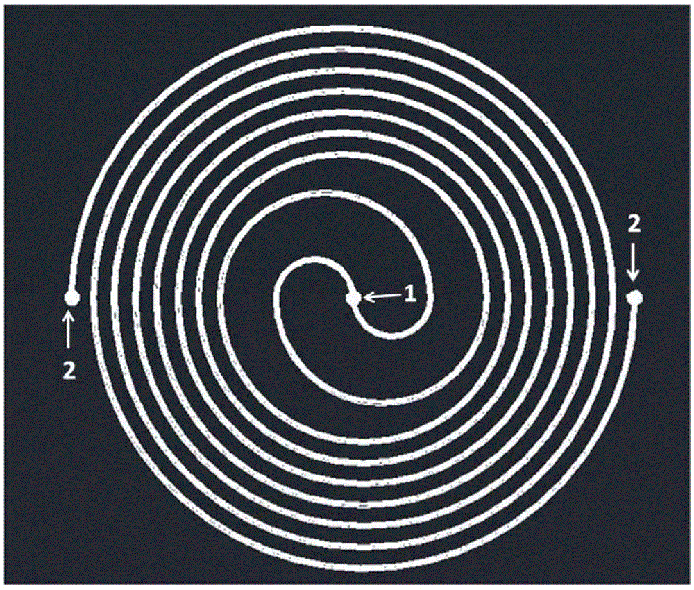
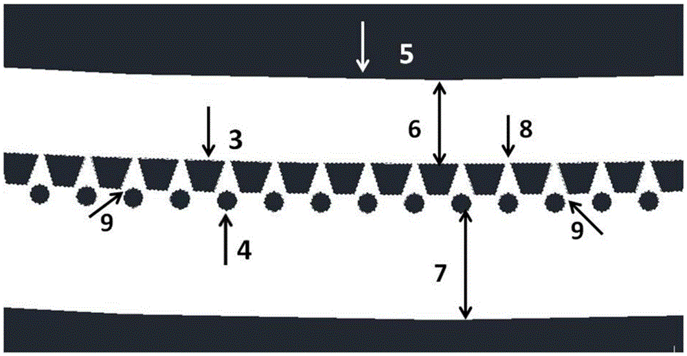
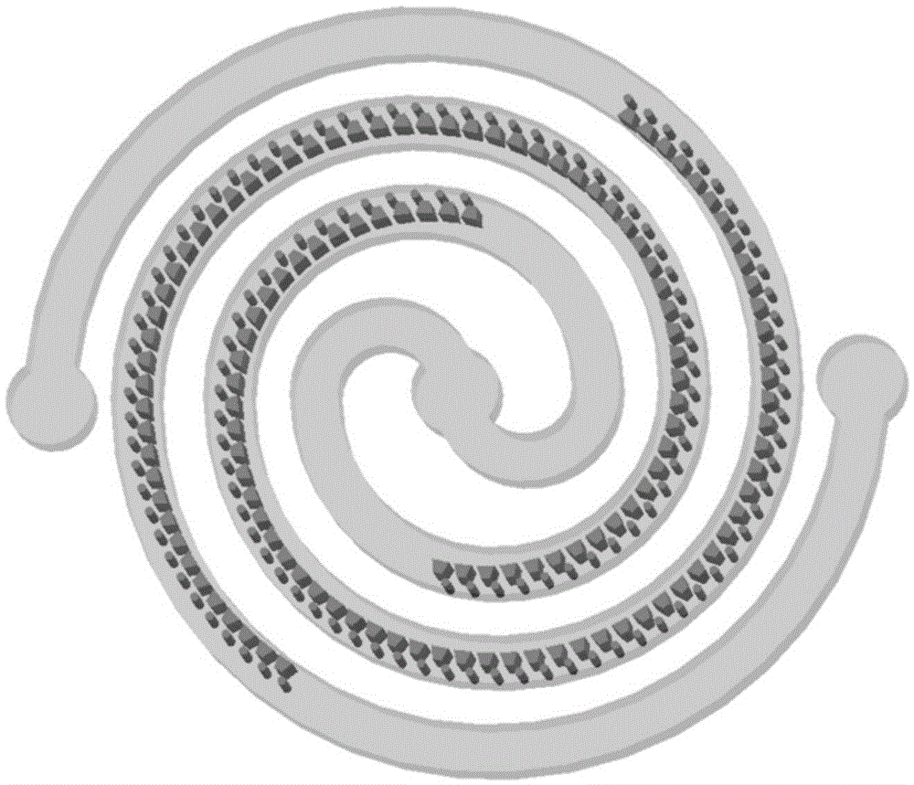
[0032]This embodiment provides a microfluidic chip that can detect 7.5ml of clinical blood samples. Each circular spiral of the chip has about 4 turns and is nested together. In the circular spiral microchannel of the microfluidic chip, there is a trapezoidal microcolumn array and a cylindrical microcolumn array parallel to the trapezoidal microcolumn array. The two bottoms of the trapezoidal microcolumn array are adjacent to each other. The distance between the corners is 5 microns. The micro-cylinder is located in the gap formed by two adjacent micro-trapezoidal upper bases, and the distance between the two corners of the two micro-trapezoidal upper bases is also 5 microns. The two layers of parallel microcolumn arrays are basically located in the middle of the microchannel. There is an inlet at the center of the connecting circle of the two circular spiral micro-column arrays, and an outlet on both sides of the left and right outer ends. Two layers of parallel microcolumn...


PUM

PropertyMeasurementUnit
sizeaaaaaaaaaa

Abstract

The invention discloses a tumor cell capturing micro-fluidic chip. The tumor cell capturing micro-fluidic chip comprises a base material and a microchannel formed inside the base material, wherein the microchannel extends along a smooth curve, an inlet and an outlet are formed in the two ends of the microchannel respectively, a microcolumn array is arranged in the vertical direction of the interior of the microchannel in a protruding mode, the microcolumn array is distributed in the extending direction of the microchannel, the end, located at the inlet, of the microcolumn array is attached to one side wall of the microchannel, and the end, located at the outlet, of the microcolumn array is attached to the other side wall of the microchannel; capture cavities with triangular cross sections are distributed in the middle of the microcolumn array at equal intervals into an array, and the three vertex angles of each capture cavity form three gaps which are 5 microns wide to be used for capturing one tumor cell 2-3 times. The chip can be used for capturing EpCAM expressed and unexpressed CTCs, EpCAM unexpressed and low-expressed CTCs and CTCs not connected with magnetic beads by means of the microcolumn array with 5 microns wide gaps.

Description

technical field [0001] The application relates to a tumor cell capture microfluidic chip and a tumor cell capture method, which can be applied to the microfluidic chip capture of various types of circulating tumor cells, and belong to the field of biophysics. Background technique [0002] Cancer, also known as malignant tumor. It is one of the leading causes of human death in the world. It is the first serious disease and chronic disease that endangers human life and health and causes death of modern humans. 90% of cancer patients die from cancer metastasis, and conquering cancer is the goal pursued by human beings. Traditional methods of surgery, chemotherapy, and radiotherapy are costly and complicated, bring great pain to patients and have recurrence. Circulating tumor cells (CTCs) are tumor cells that shed from the primary tumor and enter the blood circulation system. These tumor cells will flow to other locations to grow and develop into new tumors. The detection o...

Claims


Application Information

Patent Timeline
Patent Type & Authority Applications(China)
IPC IPC(8): C12M1/42C12M1/00C12N5/09
CPCC12M23/16C12M35/06C12N5/0693
Inventor 陈红梅聂富强窦利燕顾志鹏
Owner SUZHOU WENHAO MICROFLUIDIC TECH CO LTD
Why Eureka
Try Eureka