Quantitative method for detecting angiogenesis promotion ability of MSC

An angiogenesis and ability technology, applied in the field of stem cells, can solve the problems that hinder the clinical application of MSC and the lack of evaluation of the ability of MSC to promote angiogenesis

Inactive Publication Date: 2016-04-06
BEIJING HEALTH & BIOTECH (H&B) CO LTD
View PDF1 Cites 7 Cited by
  • Summary
  • Abstract
  • Description
  • Claims
  • Application Information

AI Technical Summary

Problems solved by technology

However, there is currently a lack of methods to evaluate the pro-angiogenic ability of

Method used


Image

  • Quantitative method for detecting angiogenesis promotion ability of MSC
  • Quantitative method for detecting angiogenesis promotion ability of MSC
  • Quantitative method for detecting angiogenesis promotion ability of MSC

Examples


Embodiment 1

[0035] Embodiment 1. Expansion and identification of HUVEC cells

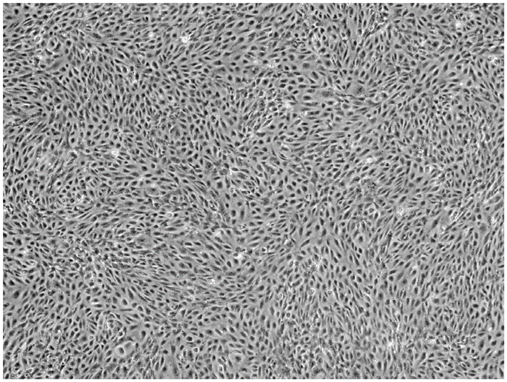
[0036] HUVEC cells were isolated from fresh umbilical cords and planted in T75 cell culture flasks at 37°C, 5% CO 2 The cells were cultured until the P5 generation, and the phenotype of the cells was identified. The results of the identification are shown in Table 1 and figure 1 As shown, the cells maintained distinct HUVEC morphology and characteristics.

[0037] Table 1. Phenotype identification of HUVEC

[0038] Umbilical vein endothelial cell markers

Embodiment 2

[0039] Example 2. Selection of CCK8 incubation time and formulation of HUVEC standard curve.

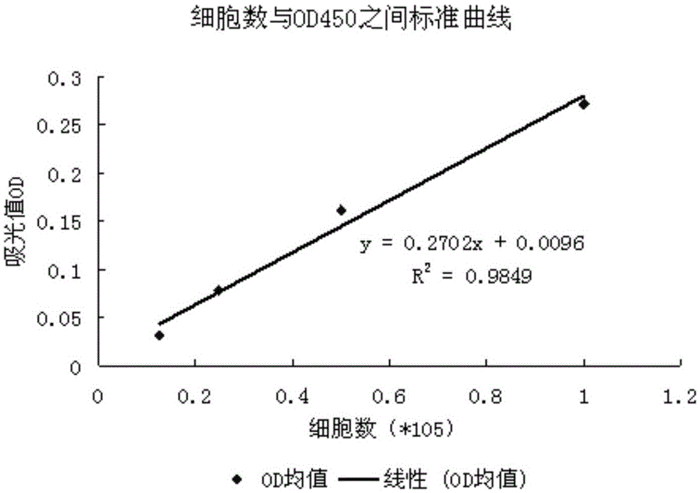
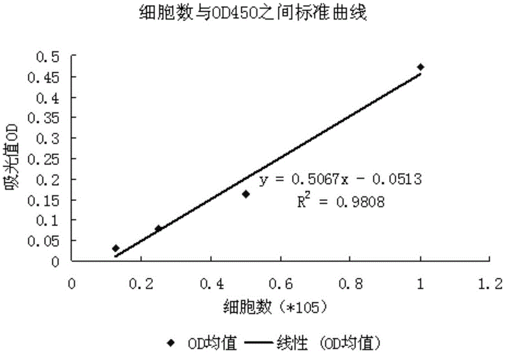
[0040] Plant HUVEC cells with different densities in 24-well plates. After adhering to the wall, add CCK8 detection solution for incubation according to the instructions of the CCK8 kit, and take part of the supernatant for enzyme labeling at 2 hours and 4 hours after incubation (OD450 / 630 ), the experimental results showed that there was a certain linear relationship between the number of viable cells detected after incubation for different times and the OD value. figure 2 is the standard curve after CCK8 incubation for 2h, image 3 is the standard curve after CCK8 incubation for 4h. With the extension of the incubation time, the OD value becomes larger, but the slope of 2hr and 4hr is the same, corresponding to the same number of cells. Finally, the incubation time was determined to be 2h, that is, the optimal incubation time was 2h.

Embodiment 3

[0041] Example 3. Selection of co-cultivation time and formulation of VEGF standard curve

[0042] HUVEC cells were planted in 24-well plates, cultured normally after starvation culture, VEGF factor was added to the culture medium, the gradient was 4ng, 2ng, 1ng, 0ng, and three replicate holes were set for each group, so as to draw a standard curve and detect the total The amount of VEGF factors secreted by MSCs into the culture medium in the culture system. Select the remaining wells plus a hanging culture dish (transwell), plant MSC in the transwell, the ratio of the number of MSC / HUVEC cells is 2 / 15, 1 / 15, 1 / 30, 1 / 60, and set 3 replicate wells for each group . The co-cultivation time was set at 48h and 72h to determine the best time for co-cultivation. Add CCK8 for incubation according to the instructions for use. After incubation for 2 hours, take part of the supernatant to measure the enzyme-labeled OD450 / 630, and analyze the number of cell proliferation and the number ...


PUM

PropertyMeasurementUnit
Gradientaaaaaaaaaa

Abstract

The invention discloses a quantitative method for detecting angiogenesis promotion ability of mesenchymal stem cells (MSC). The quantitative method is characterized by comprising the steps that a co-culture system is established, wherein MSC promote proliferation of endothelial cells by means of cytokine secretion, the proliferation number of the endothelial cells is detected, the ability of the MSC in promoting proliferation of the endothelial cells is reflected by the proliferation number of the endothelial cells, and the angiogenesis promotion ability of the MSC is quantified and evaluated. The quantitative method is suitable for detecting the angiogenesis promotion ability of MSC samples with different sources.

Description

technical field [0001] The invention relates to the field of stem cells, in particular to a quantitative method for the ability of MSCs to promote angiogenesis. Background technique [0002] Mesenchymal stem cells (MSCs) are a heterogeneous cell population derived from mesoderm, which can be obtained from most tissues of the human body. They have the ability of self-renewal, tissue repair, and immune regulation, and can differentiate into mesoderm, such as adipocytes, bone cells, chondrocytes, etc. Among them, MSCs derived from placenta and umbilical cord have great differentiation potential, strong proliferation ability, and immunogenicity. It is one of the most clinically promising pluripotent stem cells because of its low sexiness, convenient material collection, no restrictions on moral theory issues, and easy industrial preparation. [0003] Blood vessels are an important part of the human body, which constitute the main body of the circulatory system. For a long time...

Claims


Application Information

Patent Timeline
IPC IPC(8): C12Q1/06
CPCG01N33/5064G01N33/5073
Inventor 梁璐赵钦军韩之海
Owner BEIJING HEALTH & BIOTECH (H&B) CO LTD
Why Eureka
Try Eureka