System and method for three-dimensional breast X-ray and three-dimensional color Doppler ultrasound fusion imaging

An imaging system and X-ray technology, applied in the field of medical devices, can solve problems such as difficulty in seeing the complete image structure of lesion tissue, difficulty in finding details of lesions such as tiny calcifications, and breast compression

Inactive Publication Date: 2016-04-20
SINO MEDICAL DEVICE TECH
View PDF1 Cites 14 Cited by
  • Summary
  • Abstract
  • Description
  • Claims
  • Application Information

AI Technical Summary

Problems solved by technology

[0006] The main advantage of X-ray imaging is that it can identify the details of lesions such as tiny calcifications, and can detect potential breast lesions earlier; the disadvantage is that it requires a lot of compression on the breast, and it is difficult to see the complete image structure of the lesion tissue
Ultrasound imaging can clearly show the complete image structure of the diseased tissue, but it is difficult to find the details of the disease such as tiny calcifications

Method used


Image

  • System and method for three-dimensional breast X-ray and three-dimensional color Doppler ultrasound fusion imaging
  • System and method for three-dimensional breast X-ray and three-dimensional color Doppler ultrasound fusion imaging
  • System and method for three-dimensional breast X-ray and three-dimensional color Doppler ultrasound fusion imaging

Examples


Embodiment Construction

[0052] In order to have a clearer understanding of the technical features, purposes and effects of the present invention, the specific implementation manners of the present invention will now be described in detail with reference to the accompanying drawings.

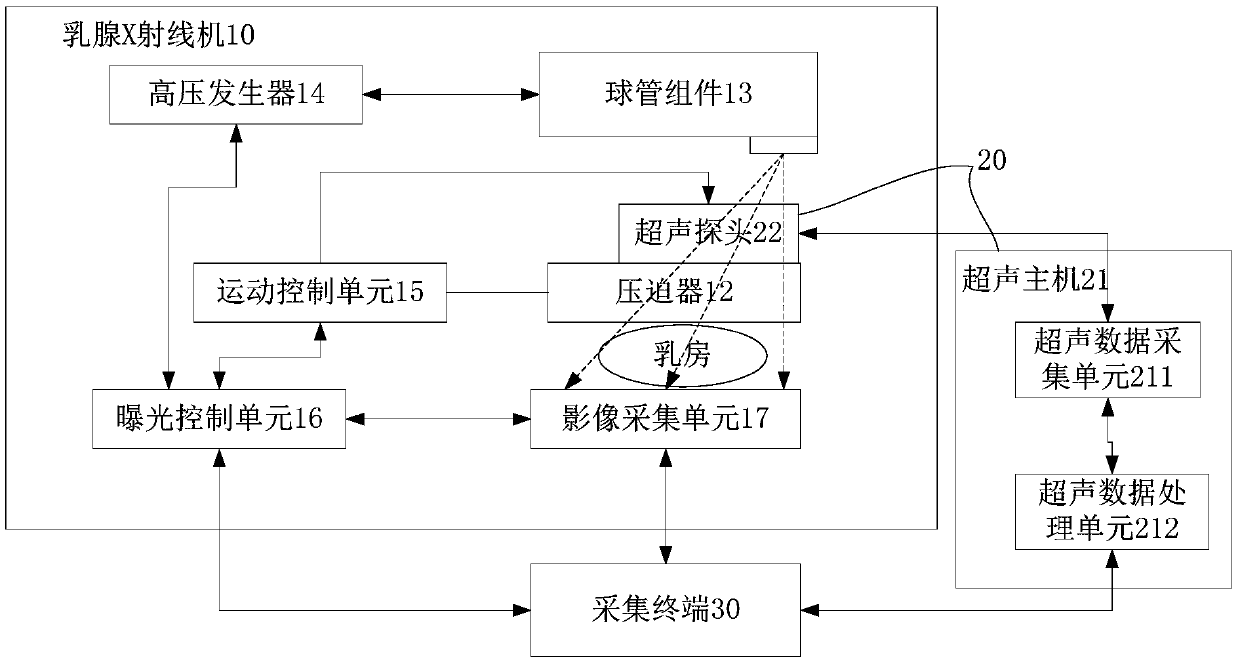
[0053] Such as figure 1 As shown, the three-dimensional breast X-ray and three-dimensional color Doppler ultrasound fusion imaging system according to an embodiment of the present invention includes a mammogram machine 10 for collecting three-dimensional X-ray images of the breast, an ultrasound machine 20 for collecting three-dimensional ultrasound images of the breast, and an image of the breast The three-dimensional X-ray image and the three-dimensional ultrasound image are fused to obtain the acquisition terminal 30 of the two-dimensional image group, and the acquisition terminal 30 is connected to the mammography machine 10 and the ultrasound machine 20 respectively. The two-dimensional image group includes two or ...


PUM


Abstract

The invention discloses a system and method for three-dimensional breast X-ray and three-dimensional color Doppler ultrasound fusion imaging. The system for fusion imaging comprises a breast X-ray machine for collecting three-dimensional X-ray images of the breast, an ultrasonic machine for collecting three-dimensional ultrasound images of the breast and a collecting terminal for performing fusion treatment on the three-dimensional X-ray images and the three-dimensional ultrasound images of the breast so as to obtain a two-dimensional image group. The two-dimensional image group comprises two or more fused two-dimensional images. The collecting terminal is in communication connection with the breast X-ray machine and the ultrasonic machine. According to the system, the advantages of the X-ray technology and the color Doppler ultrasound technology are combined, so that the breast two-dimensional image group fusing X-ray and color Doppler ultrasound imaging information is obtained, and thus clinical doctors can make a diagnosis on breast diseases more accurately and more conveniently.

Description

technical field [0001] The invention relates to the technical field of medical devices, in particular to a three-dimensional breast X-ray and three-dimensional color ultrasound fusion imaging system and method. Background technique [0002] At present, the early screening of breast cancer is mainly through mammography or ultrasound imaging. [0003] The principle of the mammary X-ray system is the same as that of other X-ray imaging systems. It mainly uses the penetrability of X-rays and the differences in the responses of different human tissues to X-rays during the process of X-rays penetrating human tissues. Images with different light and shade are formed on the image carrier. The working principle of the digital breast X-ray system is that the high-frequency pulse voltage is generated by the high-frequency and high-voltage generator through the power frequency electric energy, which is rectified and added to the X-ray tube to generate X-rays, which pass through the bre...

Claims


Application Information

Patent Timeline
Patent Type & Authority Applications(China)
IPC IPC(8): A61B6/00A61B8/08
CPCA61B6/00A61B6/466A61B6/502A61B6/5235A61B6/5247A61B8/0825A61B8/403A61B8/4416A61B8/483A61B8/5207A61B8/5238A61B8/5261
Inventor 易明军李文玉唐重陈杨旭初余兆健赖刚周人峰
Owner SINO MEDICAL DEVICE TECH
Why Eureka
Try Eureka