Fluorescent quantitative PCR detection method for detecting colonization amount of tobacco anthracnose

A fluorescence quantitative and detection method technology, applied in the field of molecular biology, can solve the problems of indistinguishability, large error, time-consuming, etc., and achieve the effects of short detection time, reduced difficulty, and simple process

Inactive Publication Date: 2016-06-08
河南省烟草公司洛阳市公司 +1
View PDF3 Cites 2 Cited by
  • Summary
  • Abstract
  • Description
  • Claims
  • Application Information

AI Technical Summary

Problems solved by technology

These methods are time-consuming and error-prone
For example, pear ringworm and pear anthracnose have simila

Method used


Image

  • Fluorescent quantitative PCR detection method for detecting colonization amount of tobacco anthracnose
  • Fluorescent quantitative PCR detection method for detecting colonization amount of tobacco anthracnose
  • Fluorescent quantitative PCR detection method for detecting colonization amount of tobacco anthracnose

Examples


Embodiment

[0040] 1. Extraction of DNA from susceptible tobacco strains

[0041] 1. Use scissors to cut the leaf tissue of the tobacco plant to an angular shape below 3mm, with a weight within the range of 10-100mg, put it into a 1.5ml Microtube, and freeze it at -20°C.

[0042] 2. Take out the frozen plant tissue and let it thaw at room temperature for about 5 minutes.

[0043]3. Gently centrifuge to collect the plant tissue at the bottom of the Microtube.

[0044] 4. Use the tip of the PipetTip to press the plant tissue to the bottom of the Microtube about 10 times for physical breaking.

[0045] 5. Add 400 μl of ExtractionSolution1 and shake vigorously for 5 seconds. If the plant tissue is still stuck at the bottom of the Microtube, flick the Microtube with your fingers to suspend it.

[0046] 6. Slightly centrifuge, add 80 μl of ExtractionSolution2, shake vigorously for 5 seconds. After adding ExtractionSolution2, a white precipitate was produced, and the solution became cloudy a...


PUM


Abstract

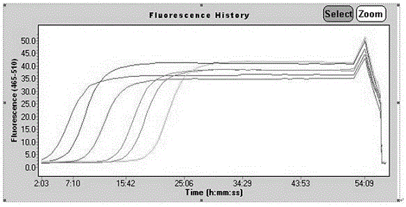
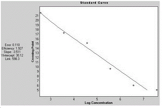
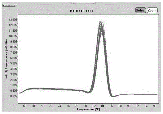
The invention relates to a fluorescent quantitative PCR detection method for detecting colonization amount of tobacco anthracnose. The method comprises the following steps: (1) preparation of a fluorescent quantitative nucleic acid amplification reaction system: a forward LNA primer, a reverse LNA primer, a template DNA and 2x fluorescent quantitative PCR reaction mixed liquor are added into a reaction tube, and 25[mu]l of ddH2O is added into the reaction system with uniformly mixing; (2) fluorescent quantitative nucleic acid amplification: the mixed liquor obtained in the step (1) is placed in a fluorescent quantitative detector for carrying out a reaction; (3) after the reaction ends, the cycle threshold C(t) of the template DNA is compared with a standard curve, and concentration of 18S ribosome RNA fragment copies in the template DNA is obtained. The method has the advantages of short detection time, high sensitivity, simple operation process, standardized operation, and high-throughput detection.

Description

technical field [0001] The invention relates to the field of molecular biology, in particular to a fluorescent quantitative PCR detection method for detecting the colonization amount of tobacco anthracnose, which is suitable for the quantitative detection of tobacco anthracnose bacteria in various susceptible tobacco strains. Background technique [0002] After tobacco anthrax was first reported by Brazil in 1922, it was also discovered in Germany, Japan, the United States, China, Australia, India, North Korea and Africa. The disease can occur in all growth stages of tobacco, but it is common and serious at the seedling stage. The leaves of seedlings are densely covered with diseased spots, and the whole tobacco seedlings are often destroyed when the disease is severe. Generally, although the seedlings are not destroyed when the disease occurs, the seedlings have poor growth potential, and they can continue to cause damage after transplanting to the field, resulting in great...

Claims


Application Information

Patent Timeline
IPC IPC(8): C12Q1/68
CPCC12Q1/6895C12Q1/686
Inventor 韦凤杰苏新宏叶红朝赵世民王惠董昆乐孔德辉李豪豪奚家勤胡利伟李芳芳赵浩宾何雷李磊王根发
Owner 河南省烟草公司洛阳市公司
Why Eureka
Try Eureka