Myocardium T1 quantifying method and device

A technology of myocardium and heart, applied in the field of myocardial T1 quantification and devices, can solve the problems of long acquisition time, many breath-hold times, low patient comfort, etc., and achieve the effect of reducing time waste and preventing deviation

Active Publication Date: 2016-06-15
SHENZHEN INST OF ADVANCED TECH
View PDF9 Cites 9 Cited by
  • Summary
  • Abstract
  • Description
  • Claims
  • Application Information

AI Technical Summary

Problems solved by technology

[0006] In the above method, each breath-hold can only collect a single-layer image, and multiple breath-holds are required to c

Method used


Image

  • Myocardium T1 quantifying method and device
  • Myocardium T1 quantifying method and device
  • Myocardium T1 quantifying method and device

Examples


Embodiment 1

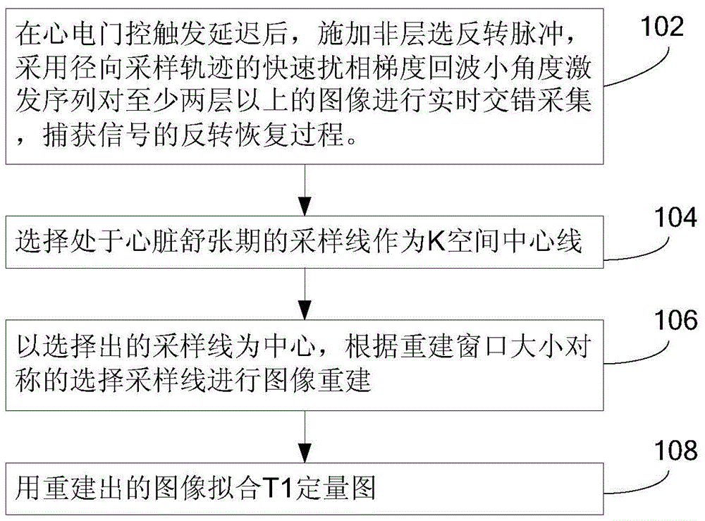
[0031] figure 1 A flowchart showing an embodiment of the method according to the present application, including:

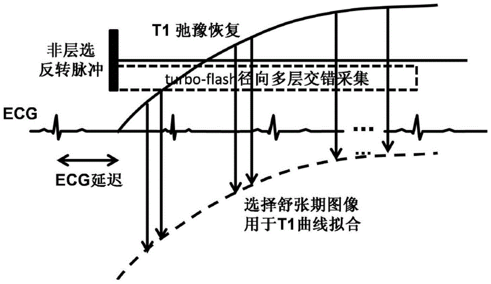
[0032] Step 102: After the ECG gating trigger delay, apply a non-slice-selective inversion pulse, and use the fast spoiler gradient echo small-angle excitation sequence of the radial sampling trajectory to perform real-time interleaved acquisition of at least two layers of images to capture the signal reverse recovery process.

[0033] Apply a non-slice-selected inversion pulse (IR), and then use the fast spoiled gradient echo small-angle excitation sequence (turbo-flash) of the radial sampling trajectory for real-time acquisition to sample the inversion recovery process of the signal, turbo- The flash adopts multi-layer interleaved acquisition mode (interleaved acquisition), and multi-layer images can be collected after one inversion pulse, such as figure 2 shown.

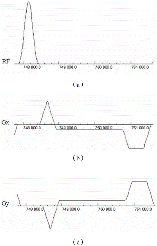
[0034] Radial sampling gradient waveform as shown in image 3 shown. In radial sampling, eac...

Embodiment 2

[0044] Figure 7 It is a schematic structural diagram of an embodiment of the device according to the present application, including: a data acquisition module, a selection module, an image reconstruction module and a fitting module.

[0045] The data acquisition module is used to apply a non-slice-selective inversion pulse after the trigger delay of the ECG gating, and use the fast spoiler gradient echo small-angle excitation sequence of the radial sampling trajectory to perform real-time interleaved acquisition of images of at least two layers, The inversion recovery process of the captured signal. One embodiment is also used to apply a non-slice-selected inversion pulse, and use the radial sampling trajectory of the fast spoiled gradient echo small-angle excitation sequence to perform real-time acquisition, and sample the inversion recovery process of the signal. The fast spoiler gradient echo small-angle excitation sequence of the radial sampling trajectory adopts a multi...


PUM


Abstract

The invention discloses a myocardium T1 quantifying method. The method includes the steps that after electrocardiograph gating trigger delay, non-layer-selection inversion pulses are applied; real-time interlaced collection of at least two layers of images is carried out with a fast spoiled gradient echo low-angle shot sequence of a radial sampling trajectory, and the inversion recovery process of signals is captured; a sampling line in diastole is selected to serve as a K space center line; with the selected sampling line as the center, the sampling line is symmetrically selected to carry out image reconstruction according to the size of a reconstruction window; a T1 quantifying graph is fitted with restructured images. The invention further discloses a device based on the method. By means of the method and the device, multiple layers of T1 quantifying images can be collected in one time of breathholding, the whole heart can be covered in two or three times of breathholding, and thus time waste and patient discomfort caused by breathholding are reduced.

Description

technical field [0001] The present application relates to the field of cardiovascular imaging, and specifically relates to a method and device for quantifying myocardial T1. Background technique [0002] Cardiovascular magnetic resonance imaging (CMR) is a non-invasive imaging technique that can evaluate the function, shape and structure of the cardiovascular system. The quantification of myocardial T1 value can be used in the diagnosis of various diseases such as myocardial infarction and myocardial fibrosis. In vivo T1 quantification of myocardium remains challenging due to acquisition time constraints imposed by heart beating and respiratory motion. [0003] The commonly used methods for measuring myocardial T1 value are as follows: [0004] (1) MOLLI (ModifiedLook-Lockerinversionrecovery): after the inversion pulse (IR), use bssfp (balancesteadystatefreeprecession) sequence imaging at different delay (TI) times, and combine the data collected multiple times into one gr...

Claims


Application Information

Patent Timeline
IPC IPC(8): A61B5/055A61B5/00
CPCA61B5/055A61B5/7207
Inventor 朱燕杰钟耀祖刘新郑海荣
Owner SHENZHEN INST OF ADVANCED TECH
Why Eureka
Try Eureka