
Skin image affected part segmentation method

A skin image and component technology, applied in the field of medical image processing, can solve problems such as segmentation interference, skin redness, and large differences in skin disease morphology and color, and achieve the effect of fast running speed and high accuracy

Inactive Publication Date: 2016-07-13
TIANJIN UNIV
View PDF3 Cites 13 Cited by
  • Summary
  • Abstract
  • Description
  • Claims
  • Application Information

AI Technical Summary

Problems solved by technology

The main difficulties at present are: (1) Because the skin is affected by many factors, the shape and color of skin diseases are quite different; (2) There are often red areas around the affected area, so that the border between the affected area and the skin is not obvious , the effect of using the gradient edge-based segmentation algorithm is not rational; (3) If there is an area other than the skin in the photo, this area may interfere with the segmentation

Method used


Image

  • Skin image affected part segmentation method
  • Skin image affected part segmentation method
  • Skin image affected part segmentation method

Examples


Embodiment Construction

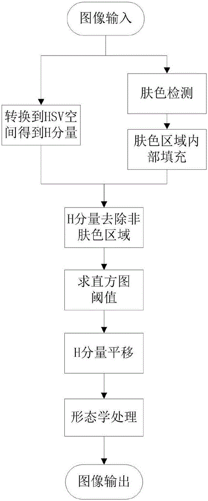
[0021] In order to further illustrate the present invention, below in conjunction with appendix figure 1 flow chart and attached Figure 2-8 The implementation example of the diagram gives a concrete example.

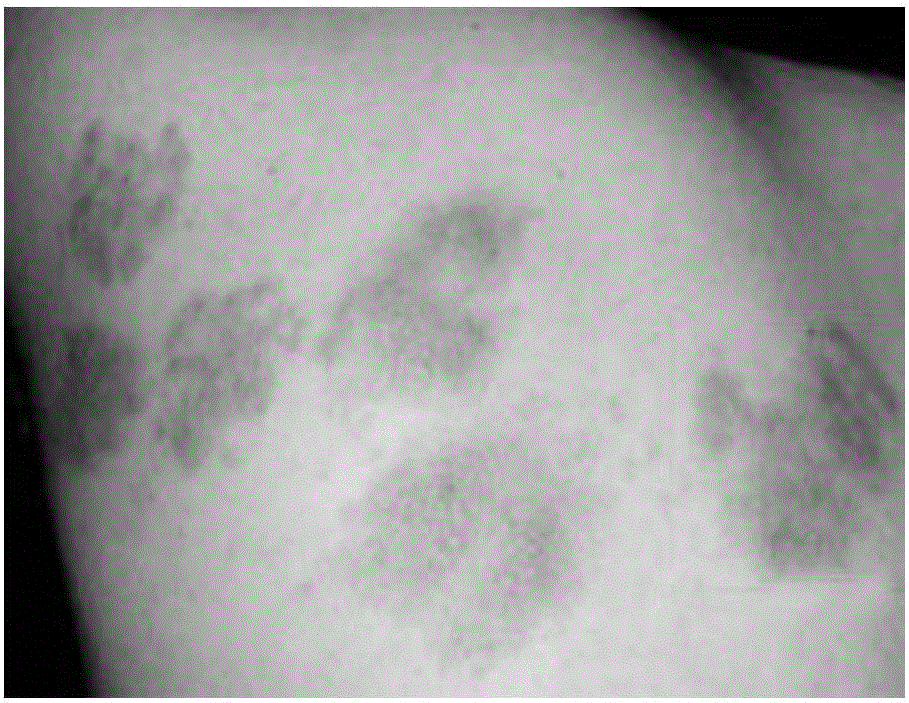
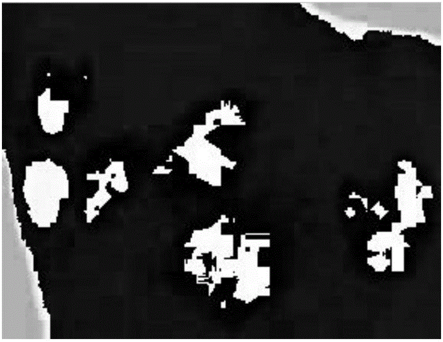
[0022] figure 2 A typical image of diseased skin is shown. There are several affected areas, in addition to two background areas outside the skin causing interference. In the picture, the boundary between some affected areas and normal skin is not obvious.

[0023] frame of reference figure 1 As shown in the flow chart, the specific skin image lesion segmentation process of the present invention is described as follows:

[0024] Step 1: Yes figure 2 The input image shown is preprocessed for denoising. Convert the image to the HSV color space to get the normalized H component map I 1 , that is, the value of the H component is between 0 and 1. The formula for calculating the normalized H component from the R, G, and B components is:

[0025] H ...


PUM


Abstract

The invention relates to a skin image affected part segmentation method comprising the steps that an input skin image is preprocessed, RGB component values are extracted, and the image is converted to an HSV color space; skin color areas and non-skin color areas are detected, and internal filling is performed in the skin color areas; pixel value histogram statistics is performed on the skin color area H component diagram obtained in 3), and a new skin color area H component diagram is obtained by using an Otsu threshold method; and binarization is performed on the skin color area H component diagram and morphological processing is performed. The skin image affected part segmentation method is suitable for segmentation of the affected part in multiple skin disease images and has the characteristics of high operation speed and high accuracy.

Description

technical field [0001] The invention relates to the field of medical image processing, in particular to a skin image lesion segmentation method. Background technique [0002] With the wide application of image processing technology in the medical field, computer diagnosis and assisted diagnosis have become a hot topic in research. Skin disease is a kind of common disease. Since the affected area can be directly observed, computer diagnosis and analysis of skin disease can be realized through image processing technology. The first step in the computer diagnosis of skin diseases is to segment the affected area of ​​the skin disease from the collected skin pictures. Only when the accurate and efficient segmentation of the affected area is realized can the various characteristics of the affected area be further analyzed. The main difficulties at present are: (1) Because the skin is affected by many factors, the shape and color of skin diseases are quite different; (2) There are...

Claims


Application Information

Patent Timeline
IPC IPC(8): G06T7/00G06T7/40
CPCG06T2207/30088
Inventor 王昇刘开华马永涛
Owner TIANJIN UNIV
Features
  • Generate Ideas
  • Intellectual Property
  • Life Sciences
  • Materials
  • Tech Scout
  • Unparalleled Data Quality
  • Higher Quality Content
  • 60% Fewer Hallucinations
Learn More