Method for measuring diameter of maximum choroid blood vessel based on image segmentation

A technology of image segmentation and measurement method, applied in the field of measurement, can solve the problems of non-perpendicular measurement of blood vessel diameter, easy to cause errors, lack of accurate objective quantitative detection, etc., to achieve the effect of improving measurement accuracy and efficiency and reducing measurement errors

Inactive Publication Date: 2016-07-20
CAPITAL UNIVERSITY OF MEDICAL SCIENCES
View PDF2 Cites 4 Cited by
  • Summary
  • Abstract
  • Description
  • Claims
  • Application Information

AI Technical Summary

Problems solved by technology

In recent years, due to the incomplete and unclear performance of choroidal vessels in frequency-domain optical coherence tomography (SD-OCT) images, the methods for monitoring choroidal vessels are limited, and the lack of accurate and objective quantitative detection tools is often used clinically. The disadvantage is that the retinal blood vessels are not completely regular and radially distributed with the optic disc as the center, so when scanning the blood vessels within the concentric circle with the optic disc as the center, there will be non-perpendicular measurement of the blood vessel diameter, resulting in errors;
Therefore, the traditional target extraction method is difficult to effectively give the choroidal vessel area
In order to overcome the low accuracy of existing methods for measuring choroidal blood vessels and easily lead to errors, the present invention provides a method with high accuracy and capable of obtaining the largest diameter of choroidal blood vessels

Method used


Image

  • Method for measuring diameter of maximum choroid blood vessel based on image segmentation
  • Method for measuring diameter of maximum choroid blood vessel based on image segmentation
  • Method for measuring diameter of maximum choroid blood vessel based on image segmentation

Examples


Embodiment Construction

[0019] refer to Figure 1-4 , to further illustrate the present invention:

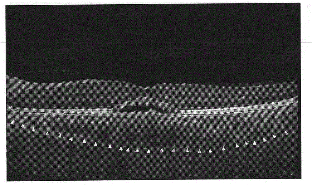
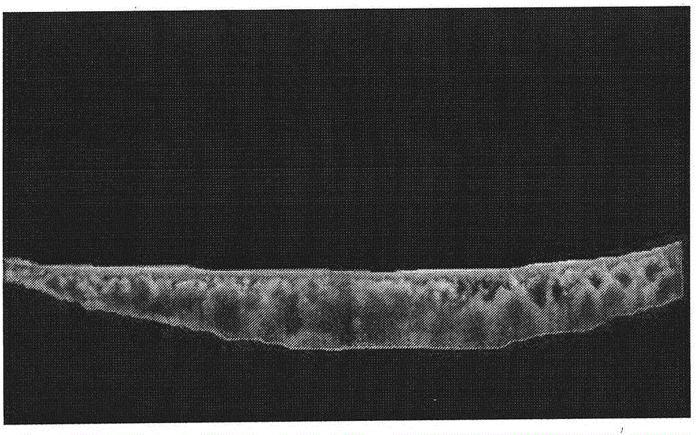
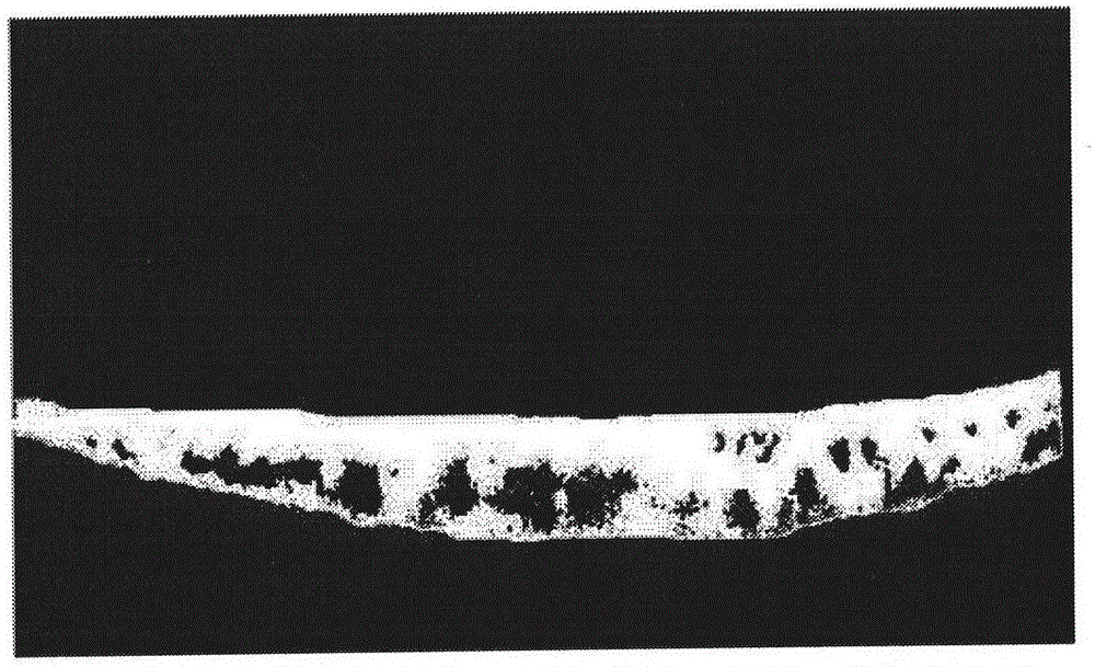
[0020] A method for measuring the diameter of the largest vessel in the choroid based on image segmentation, firstly obtain SD-OCT retinal images such as figure 1 As shown, the choroidal layer of the acquired SD-OCT retinal image is measured by image segmentation method, including image preprocessing module, image segmentation module and measurement result output module; wherein the image preprocessing module includes choroidal layer image filtering and choroidal layer image enhancement; the image segmentation module is used to segment the choroidal layer using an image segmentation algorithm, such as figure 2 As shown, and segment the largest vessel in the choroid layer, as image 3 shown, and then calculate the value of the divided area; the measurement result output module is used to output the measurement result.

[0021] The measurement method consists of the following steps:

[0022] Step 1...


PUM


Abstract

The invention discloses a method for measuring the diameter of the maximum choroid blood vessel based on image segmentation. The method comprises the following steps: firstly performing image pre-processing on a choroid in a SD-OCT retina image, then adopting the method of image segmentation to extract a region of interest and performing related calculation on the region, and finally outputting a measuring result. According to the invention, the method reduces measuring errors by making the obtained diameter of the choroid maximum blood vessel with an improved precision than the diameter obtained by manual measuring. The method, by using the simple and rapid image segmentation technology, increases accuracy and efficiency in measuring the diameter of the choroid blood vessel, and has great significance in facilitating successive choroid diseases analysis and improving doctor's working efficiency.

Description

technical field [0001] The invention relates to a measurement method, in particular to a method for measuring the maximum choroidal vessel diameter based on image segmentation. Background technique [0002] The choroid layer is composed of a large number of blood vessels, which provide nutrition for the retinal layer and is closely related to retinal diseases. Abnormal changes such as choroidal vasodilation, congestion, and high permeability can cause choroid-related fundus diseases. The morphology of the choroid plays an irreplaceable role in auxiliary diagnosis in clinical practice. concern of ophthalmology clinicians. In recent years, due to the incomplete and unclear performance of choroidal vessels in frequency-domain optical coherence tomography (SD-OCT) images, the methods for monitoring choroidal vessels are limited, and the lack of accurate and objective quantitative detection tools is often used clinically. The disadvantage is that the retinal blood vessels are n...

Claims


Application Information

Patent Timeline
Patent Type & Authority Applications(China)
IPC IPC(8): G06T7/00
CPCG06T7/0012G06T2207/20182G06T2207/30041G06T2207/30101
Inventor 刘加峰张海燕
Owner CAPITAL UNIVERSITY OF MEDICAL SCIENCES
Why Eureka
Try Eureka