Mark-method-based B ultrasound imaging and microwave imaging fused method and system

A technology of microwave imaging and labeling, which is used in image enhancement, image analysis, image data processing, etc.

Inactive Publication Date: 2016-09-28
YITI INTELLIGENT TECH LTD SHENZHEN CITY
View PDF6 Cites 0 Cited by
  • Summary
  • Abstract
  • Description
  • Claims
  • Application Information

AI Technical Summary

Problems solved by technology

[0005] The technical problem solved by the present invention is to construct a method and system based on the labeling method for fused B-ultrasound imaging and microwave imaging, and overcome the technical problem that a single detection device in the prior art cannot simultaneously utilize two images for complementary advantages

Method used


Image

  • Mark-method-based B ultrasound imaging and microwave imaging fused method and system
  • Mark-method-based B ultrasound imaging and microwave imaging fused method and system
  • Mark-method-based B ultrasound imaging and microwave imaging fused method and system

Examples


Embodiment Construction

[0021] The technical solutions of the present invention will be further described below in conjunction with specific embodiments.

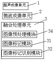
[0022] Such as figure 1 Shown, the specific embodiment of the present invention is: provide a kind of method based on the fusion B ultrasonic imaging of labeling method and microwave imaging, comprise the steps:

[0023] Separate imaging: B-ultrasound signals are generated in the area to be tested, and the echo signals of the B-ultrasound signals are received for B-ultrasound imaging; microwave signals are generated in the area to be tested, and the echo signals of the microwave signals are received for microwave imaging;

[0024] The specific implementation process is as follows: a B-ultrasound image is generated according to the received B-ultrasound signal, and a microwave image is generated according to the received microwave echo signal.

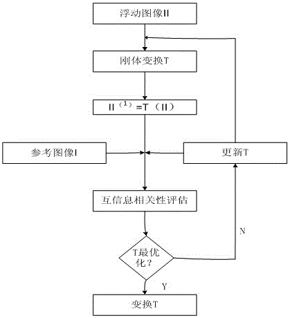
[0025] Image fusion: using the microwave image as a reference image, registering the B-ultrasound image...


PUM


Abstract

The present invention relates to a method and system for fusing B-ultrasound imaging and microwave imaging based on a marking method. By mapping the pixel points of one image in the B-ultrasound image and the microwave image to the other image, the two The fusion of the two images is completed when the relevant pixels of the two images are consistent in spatial position. In the process of diagnosing early cancer patients, different imaging modalities can be used to complement each other. The potential of image fusion lies in the comprehensive application of information obtained from these imaging devices, which can accurately determine the spatial location, geometry, and relationship between the lesion and its surroundings. The spatial relationship between biological tissues can be used to diagnose diseases in a timely and efficient manner. It can also be used to formulate surgical plans, track pathological changes, and evaluate therapeutic effects.

Description

technical field [0001] The invention relates to an image fusion method and system, in particular to a B-ultrasound image and microwave image fusion method and system. Background technique [0002] Microwave imaging determines the positional distribution of the dielectric constant and conductivity in the living body, and processes the measurement data to find out those tissues in the imaged body whose dielectric constant is different from the normal value. Its reliability has been tested in various studies. Confirmation. Existing microwave imaging systems mainly focus on imaging algorithms and antenna design. Existing microwave imaging algorithms mainly include ultra-wideband radar imaging and microwave tomography. Ultra-wideband radar imaging needs to reconstruct the The spatial distribution of scattering intensity can process signals in several GHz frequency bands and effectively improve the spatial resolution of the imaging system. Microwave tomography requires reconstruct...

Claims


Application Information

Patent Timeline
IPC IPC(8): G06T7/00
CPCG06T7/0012G06T2207/30096
Inventor 罗林
Owner YITI INTELLIGENT TECH LTD SHENZHEN CITY
Why Eureka
Try Eureka