
Robotic control of surgical instrument visibility

A technology of robots and instruments, applied in the field of robot control

Active Publication Date: 2016-10-05
KONINKLJIJKE PHILIPS NV
View PDF5 Cites 2 Cited by
  • Summary
  • Abstract
  • Description
  • Claims
  • Application Information

AI Technical Summary

Problems solved by technology

In practice, two problems usually arise: first, the surgeon usually moves one or both instruments out of the view of the endoscope
However, calibration of endoscopes is a technical challenge for medical personnel and should preferably be avoided

Method used


Image

  • Robotic control of surgical instrument visibility
  • Robotic control of surgical instrument visibility
  • Robotic control of surgical instrument visibility

Examples


Embodiment Construction

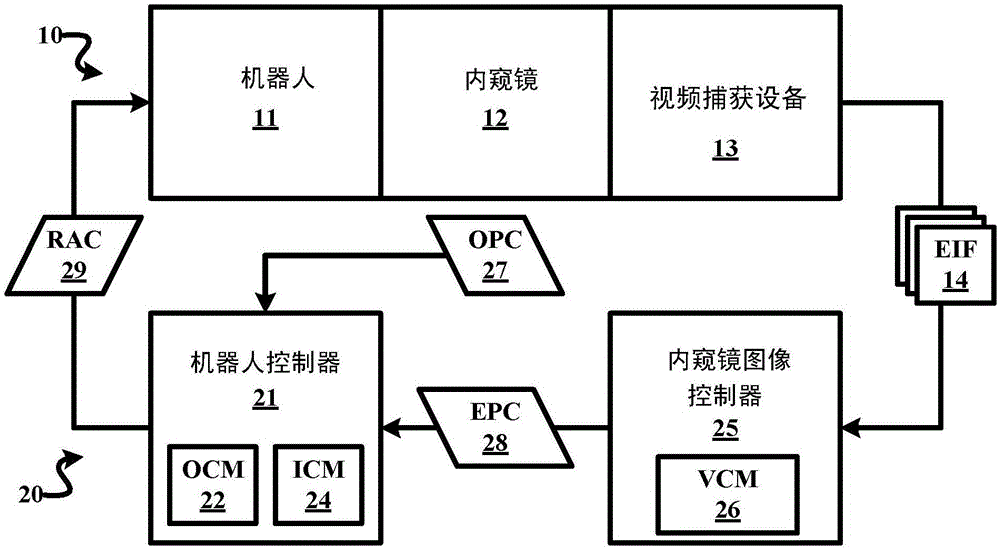
[0020] Such as figure 2 As shown, the robotic guidance system employs a robotic unit 10 and a control unit 20 for any endoscopic procedure involving endoscopic imaging of an anatomical region (eg, cranial region, thoracic region, abdominal region, patella region, etc.). Examples of such endoscopic procedures include, but are not limited to, minimally invasive cardiac surgery (e.g., coronary artery bypass grafting or mitral valve replacement), laparoscopic surgery (e.g., hysterectomy, prostatectomy, and gallbladder surgery), Natural Oral Endoscopic Surgery (NOTES), Single Incision Laparoscopic Surgery (SILS), Lung / Bronchoscopic Surgery, and Minimally Invasive Diagnostic Interventions (eg, Arthroscopy).

[0021] The robotic unit 10 comprises a robot 11 , an endoscope 12 rigidly attached to the robot 11 and a video capture device 13 attached to the endoscope 12 .

[0022] Herein, a robot 11 is broadly defined as any robotic device structurally configured with motorized control f...


PUM


Abstract

A robot guiding system employs a robot unit (10) including an endoscope (12) and a robot (11), and a control unit (20) including an endoscopic image controller (22) and a robot controller (21). In operation, the endoscope (12) generates an endoscopic image of an anatomical region as the robot (11) move the endoscope (12) within the anatomical region in response to robot actuator commands. The endoscopic image controller (22) controls a display of the endoscopic image(14) of the anatomical region and generates endoscope pose commands to maintain a visibility of two or more interventional instruments within the display of the endoscopic image(14) relative to a center of the endoscopic image (14). The robot controller (21) generates the robotic actuator commands responsive to the endoscope pose commands.

Description

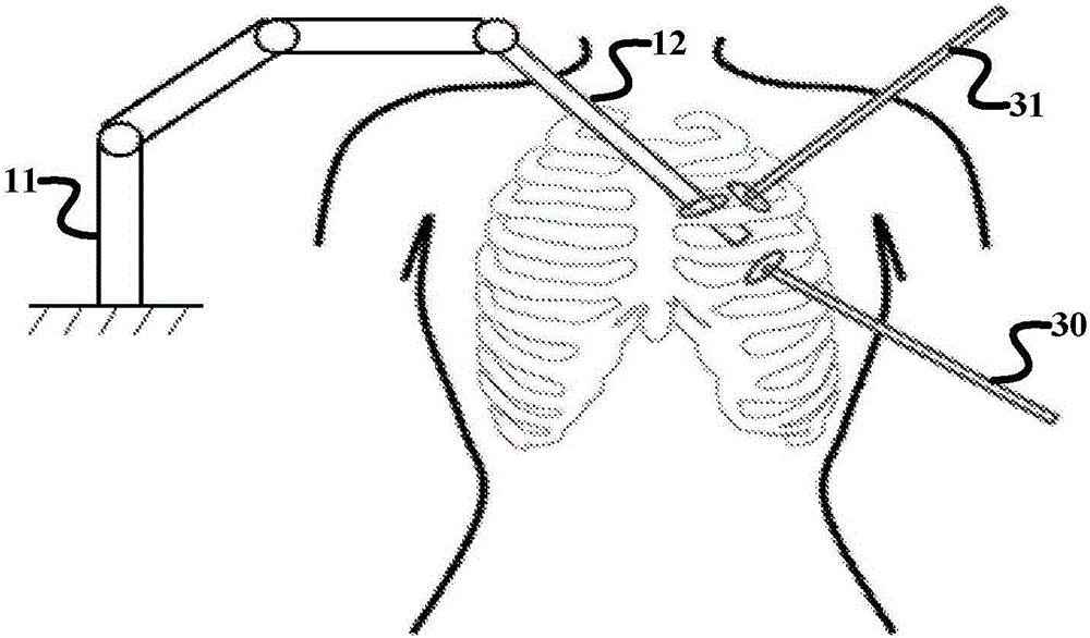
technical field [0001] The present invention generally relates to robotic control of endoscopes during minimally invasive surgical procedures (eg, minimally invasive coronary artery bypass graft surgery). The invention specifically relates to maintaining visibility of surgical instruments within an endoscopic image. Background technique [0002] Minimally invasive procedures are performed using elongated instruments that are inserted into the patient's body through small ports. An endoscopic camera is also inserted into the patient via a small port to provide visualization of the instruments relative to the surgical site. For example, FIG. 1 shows the insertion of an elongated instrument 30 through a small port into the patient's body and the endoscopic camera 12 also inserted through the small port to provide visualization of the instrument 30 relative to the surgical site. [0003] Currently, the surgeon holds and manually controls two (2) surgical instruments during sur...

Claims


Application Information

Patent Timeline
IPC IPC(8): A61B34/20A61B34/30A61B17/00
CPCA61B17/00234A61B2576/00A61B2034/2065A61B1/00006A61B1/00133A61B34/30A61B1/000094A61B2034/2057A61B2034/301A61B1/00045A61B1/00149A61B1/045A61B1/3132
Inventor A·波波维奇D·P·努南
Owner KONINKLJIJKE PHILIPS NV
Learn More