Automatic identification method of microaneurysm in fundus colorful image

An automatic recognition and fundus image technology, which is applied in the field of image processing, can solve problems such as difficult identification and small microaneurysms, and achieve the effects of improving work efficiency, improving image quality, and enhancing features

Active Publication Date: 2016-11-09
SHANGHAI FIRST PEOPLES HOSPITAL +1
View PDF5 Cites 11 Cited by
  • Summary
  • Abstract
  • Description
  • Claims
  • Application Information

AI Technical Summary

Problems solved by technology

[0004] The technical problem to be solved by the present invention is to propose an automatic recognition method for microaneurysms, which can achieve precise positioning and segmentation of microaneurysms, in view of the characteristics of microaneurysms in color images of ...

Method used


Image

  • Automatic identification method of microaneurysm in fundus colorful image
  • Automatic identification method of microaneurysm in fundus colorful image
  • Automatic identification method of microaneurysm in fundus colorful image

Examples


Embodiment Construction

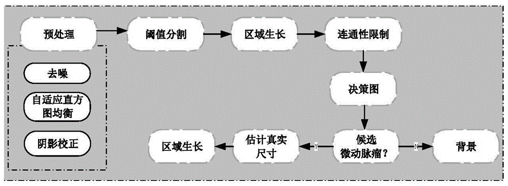
[0023] figure 1 It is a schematic diagram of automatic recognition of microaneurysms in an embodiment of the present invention. Such as figure 1 As shown, first select the green channel image in the RGB three channels, because the red lesion in the green channel image has the highest contrast with other tissues. Use median filtering technology to eliminate image noise, and use adaptive histogram equalization technology to enhance the contrast of the image. After the histogram is equalized, the shadow area caused by the non-uniform illumination will be enhanced. In order to effectively remove the influence of the shadow, the shadow correction technology is used to remove the slow gradient change in the background. In actual processing, the shading correction is to subtract the original image from the image after the median filtering process, so as to obtain the shadow-corrected image, and complete the entire preprocessing process.

[0024] figure 2 It is a schematic diagram o...


PUM


Abstract

The invention discloses an automatic identification method of microaneurysm in a fundus colorful image. The method comprises the following steps: firstly, carrying out preprocessing and shade correction on an image of a green channel, then, utilizing the local information of a sliding window to determine an adaptive threshold value to carry out binarization on the image, obtaining a decision graph through a double-window filter based on connectivity restriction, and carrying out region growing on the decision graph, wherein the result of the region growing is the accurate identification microaneurysm. By use of the whole method, the fine identification and segmentation of the microaneurysm can be realized, identification missing and misjudgement can be effectively avoided, the processes of feature extraction and classifier classification do not need to be adopted, and identification efficiency is effectively improved.

Description

Technical field [0001] The invention belongs to the field of image processing, and specifically relates to a method for automatically identifying microaneurysms in fundus images based on connectivity limitations. Background technique [0002] A microaneurysm is a sac-like swelling located in the wall of a capillary vessel. It is usually a round, discrete, small area of ​​dark red lesions. Because microaneurysms are about 10-100μm in diameter, they are difficult to detect with the naked eye, and doctors have a heavy workload and low efficiency. And because of its small surface, high similarity to blood vessels, and low background contrast, traditional automatic recognition methods are also difficult to effectively recognize. [0003] At present, the microaneurysm recognition methods proposed are mostly based on information such as the shape and color of the microaneurysm. The candidate microaneurysm points are selected through morphology and matched filtering, and then the relevant...

Claims


Application Information

Patent Timeline
IPC IPC(8): G06K9/00
CPCG06T5/10G06T2207/20032G06T2207/10024G06T2207/20182G06T2207/30041G06T2207/30096G06V10/255G06V10/30G06V2201/032G06F18/00
Inventor 许迅杨杰余奇刘梦雪
Owner SHANGHAI FIRST PEOPLES HOSPITAL
Why Eureka
Try Eureka