Ultrasound gray-scale imaging system and method

An imaging system, ultrasonic image technology, applied in radio wave measurement system, ultrasonic/sonic/infrasonic diagnosis, sonic diagnosis, etc., to achieve the effect of solving distortion and improving time resolution

Active Publication Date: 2016-11-09
SHENZHEN MINDRAY BIO MEDICAL ELECTRONICS CO LTD
View PDF6 Cites 24 Cited by
  • Summary
  • Abstract
  • Description
  • Claims
  • Application Information

AI Technical Summary

Problems solved by technology

[0004] Based on this, it is necessary to provide an ultrasonic gray-scale imaging system and method for the distortion of traditional gray-scale blood flow imaging when displaying high-speed blood flow.

Method used


Image

  • Ultrasound gray-scale imaging system and method
  • Ultrasound gray-scale imaging system and method
  • Ultrasound gray-scale imaging system and method

Examples


Embodiment Construction

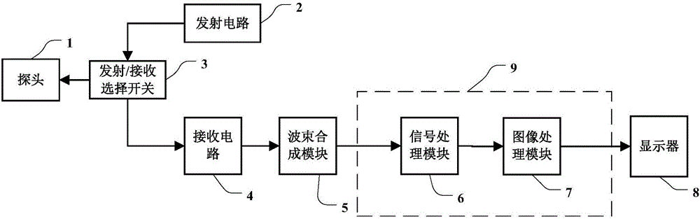
[0036] Figure 1a It is a schematic structural block diagram of a B-mode ultrasonic gray-scale imaging system according to an embodiment of the present invention. Such as Figure 1a As shown, the ultrasonic grayscale imaging system includes: a probe 1 , a transmitting circuit 2 , a transmitting / receiving selection switch 3 , a receiving circuit 4 , a beamforming module 5 , a signal processing module 6 , an image processing module 7 , and a display 8 . The above-mentioned signal processing module 6 and image processing module 7 may be implemented based on one or more processors. During the ultrasonic imaging process, the transmitting circuit 2 transmits the delayed-focused transmitting pulse with a certain amplitude and polarity to the probe 1 through the transmitting / receiving selection switch 3 . Probe 1 is excited by the transmitted pulse, transmits the ultrasonic beam to the scanning target containing the fluid, receives the ultrasonic echo with fluid information reflected ...


PUM


Abstract

An ultrasound gray-scale imaging system and method. The system comprises a probe (1); a transmission circuit (2) for exciting the probe (1) to transmit non-focused ultrasound beams to fluid; a receiving circuit (4) and a beam-forming module (5) for receiving echoes of the non-focused ultrasound beams and obtaining non-focused ultrasound echo signals; a signal processing module (6) for obtaining fluid display data after performing signal detection and enhancement process on the multi-group of non-focused ultrasound echo signals; an image processing module (7) for performing data conversion on the fluid display data and obtaining a B mode ultrasound image sequence; and a display (8) for displaying the B mode ultrasound image sequence to present the visual effect of fluid dynamic flow. A plurality of scanning lines even the whole image can be obtained for one time by utilization of non-focused wave or plane wave emission, time resolution of the ultrasound image can be largely improved, and the distortion problem when high-speed blood flow is displayed by traditional gray-scale blood flow imaging is solved.

Description

technical field [0001] The present invention relates to an ultrasonic imaging system, and in particular to an ultrasonic blood flow imaging method which uses a gray scale image to directly display blood flow. Background technique [0002] Gray-scale blood flow imaging is an ultrasonic blood flow imaging method that directly displays the blood flow by using a gray-scale image after processing the obtained ordinary B-image signal. Traditional gray-scale blood flow imaging uses focused wave emission and line-by-line scanning, so that the accuracy and sensitivity of the blood flow obtained in this way are high. The flow signal in the blood vessel is enhanced through wall filtering, and then combined with the tissue signal to combine the blood vessel wall and The flow of red blood cells in blood vessels is simultaneously displayed in grayscale images. The dynamic image of gray-scale blood flow imaging can intuitively show the difference between flowing blood and non-flowing tiss...

Claims


Application Information

Patent Timeline
Patent Type & Authority Applications(China)
IPC IPC(8): A61B8/06
CPCA61B8/06A61B6/547G01S15/8927G01S15/8979A61B8/461A61B8/5207A61B8/5246G01S15/8906
Inventor 杜宜纲
Owner SHENZHEN MINDRAY BIO MEDICAL ELECTRONICS CO LTD
Why Eureka
Try Eureka