Automatic segmentation system and method for digestive tract internal image

An automatic segmentation and digestive tract technology, applied in image analysis, image enhancement, image data processing, etc., can solve the problems of insufficient utilization and poor effect

Active Publication Date: 2016-12-07
安翰科技(武汉)股份有限公司
View PDF1 Cites 21 Cited by
  • Summary
  • Abstract
  • Description
  • Claims
  • Application Information

AI Technical Summary

Problems solved by technology

In the first stage of this method, only the first and second moments of the color are used, and other image information is not fully utilized

Method used


Image

  • Automatic segmentation system and method for digestive tract internal image
  • Automatic segmentation system and method for digestive tract internal image
  • Automatic segmentation system and method for digestive tract internal image

Examples


Embodiment Construction

[0046] The present invention will be further described in detail below in conjunction with the drawings and specific embodiments:

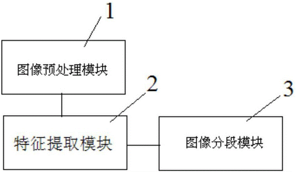
[0047] An automatic segmentation system for images in the digestive tract, such as figure 1 Said, it includes an image preprocessing module 1, a feature extraction module 2 and an image segmentation module 3. The signal output terminal of the image preprocessing module 1 is connected to the signal input terminal of the feature extraction module 2, and the signal output of the feature extraction module 2 Terminal is connected to the signal input terminal of the image segmentation module 3;

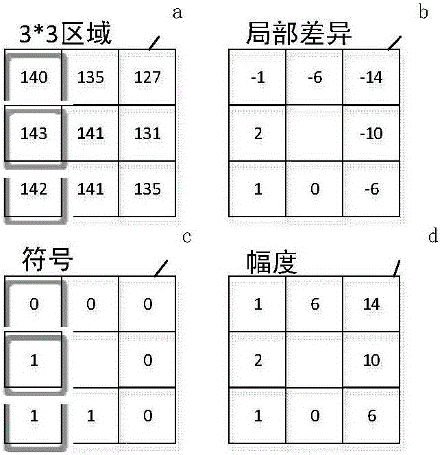
[0048] The image preprocessing module 1 is used to exclude invalid images whose grayscale average exceeds a preset threshold, and remove invalid areas in the image to reduce the images that need to be processed. The invalid areas in the image include food residue areas, bubble areas, and mucus areas. And the area in the image where the mean gray value exceeds the p...


PUM


Abstract

The invention discloses an automatic segmentation system for a digestive tract internal image. The system comprises an image preprocessing module, a feature extraction module and an image segmentation module, wherein the signal output end of the image preprocessing module is connected with the signal input end of the feature extraction module, and the signal output end of the feature extraction module is connected with the signal input end of the image segmentation module. Through the automatic segmentation system, the time for a doctor to search for images of different parts of the digestive tract can be shortened, the workload is reduced, and work efficiency is improved.

Description

Technical field [0001] The present invention relates to the technical field of computer-aided detection, in particular to an automatic segmentation system and method for images in the digestive tract. technical background [0002] The wireless capsule endoscope is a miniature camera similar in size and shape to the ordinary capsule. It enters the digestive tract through oral administration. The peristalsis of the digestive tract powers it to move in the digestive tract. The capsule endoscope takes images of the digestive tract and It is transmitted to the outside of the body through a wireless transmission system for display. [0003] The digestive tract can be divided into esophagus, stomach, small intestine, colon, etc. Each part has its own unique diseases, such as reflux esophagitis in the esophagus, antral inflammatory hyperplasia in the stomach, Crohn's disease in the small intestine, etc. . An experienced reading doctor needs to spend more time to distinguish and classify ...

Claims


Application Information

Patent Timeline
IPC IPC(8): G06T7/00G06K9/62G06F19/00
CPCG16H50/20G06T2207/30004G06F18/2411
Inventor 袁文金张皓张行王新宏段晓东肖国华
Owner 安翰科技(武汉)股份有限公司
Why Eureka
Try Eureka