A Segmentation Method of Cranial Magnetic Resonance Image Based on Spatial Mixture Model

A magnetic resonance image and space mixing technology, applied in image analysis, image enhancement, image data processing, etc., can solve the problems of noise influence, large amount of calculation, and large algorithm complexity, etc., and achieve strong noise resistance and accurate model solution Efficient effect

Active Publication Date: 2020-03-10
HUAQIAO UNIVERSITY
View PDF2 Cites 0 Cited by
  • Summary
  • Abstract
  • Description
  • Claims
  • Application Information

AI Technical Summary

Problems solved by technology

Compared with GMM, this method has stronger robustness, so the segmentation effect is further improved, but the algorithm complexity is larger
K. Blekas et al. (K. Blekas, A. Likas, N.P. Galatsanos, and I.E. Lagaris, "A spatially constrained mixture model for image segmentation", IEEE Trans. Neural Netw., vol. 16, no. 2, pp. 494– 498, 2005) proposed a spatially varying finite mixture model (SVFMM), which uses Gibbs sampling and uses the maximum posterior probability for parameter estimation, but this method requires additional calculation steps to ensure that the mixture coefficient of the mixture model satisfies The condition that the value is positive and the sum is one, thus resulting in the disadvantages of high model complexity and large amount of calculation
[0005] Generally speaking, in the segmentation of cranial magnetic resonance images, it is necessary to solve the two major problems of large amount of calculation and noise influence. The former belongs to the problem of space and time overhead, and the latter belongs to the problem of accuracy.

Method used


Image

  • A Segmentation Method of Cranial Magnetic Resonance Image Based on Spatial Mixture Model
  • A Segmentation Method of Cranial Magnetic Resonance Image Based on Spatial Mixture Model
  • A Segmentation Method of Cranial Magnetic Resonance Image Based on Spatial Mixture Model

Examples


Embodiment Construction

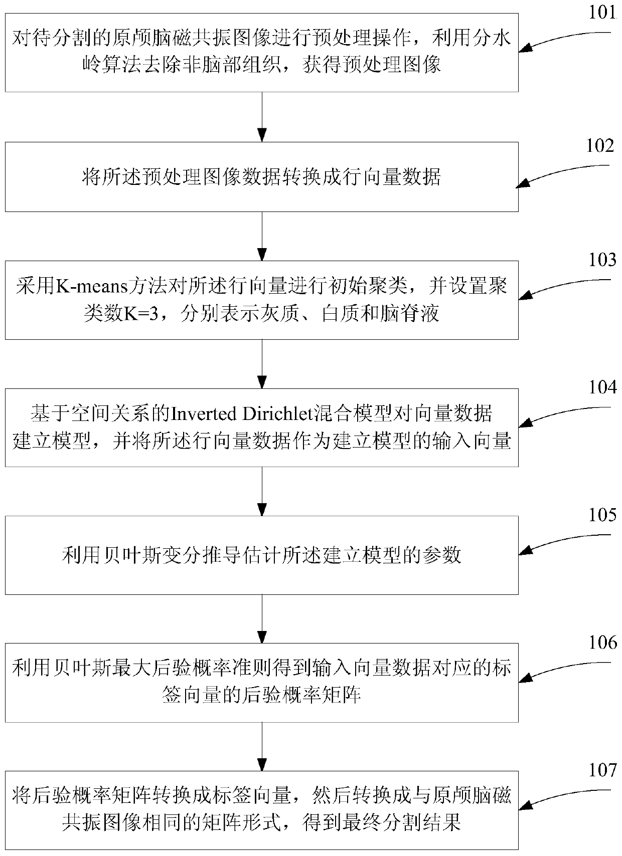
[0033] Such as figure 1 As shown, a brain MRI image segmentation method based on the spatial mixture model, the steps are as follows:

[0034] Step 101, performing a preprocessing operation on the original brain magnetic resonance image to be segmented: using the watershed algorithm to remove non-brain tissue, and obtaining a preprocessed image with N rows and M columns;

[0035] Step 102, converting the preprocessed image into a row vector;

[0036] Specifically, follow the steps below to convert the preprocessed image into a row vector:

[0037] First read the preprocessed image matrix in the form of columns to obtain an N*M-dimensional column vector;

[0038] Transpose an N*M-dimensional column vector into a corresponding row vector.

[0039] Step 103, using the K-means method to initially cluster the row vectors, and setting the number of clusters K=3, respectively representing gray matter, white matter and cerebrospinal fluid;

[0040] Step 104, building a model for t...


PUM


Abstract

The invention discloses a brain magnetic resonance image segmentation method based on a mixed space model. The method comprises steps that an inputted brain magnetic resonance image is pre-processed to acquire an image without brain tissues; the image data after pre-processing is converted into a row vector taken as an input vector; modeling for the image data after vectorization is carried out by utilizing an Inverted Dirichlet mixed model based on space relationship, a k-means method is further employed to carry out model initiation; relevant parameters of a Bayes variation derivation technology estimation mixed model are employed; a belonging category of each pixel point is determined through calculating posterior probability, and a new label vector is acquired; the label vector is taken as an output vector and is converted as a gray matrix, and a segmentation result is finally acquired. The method is advantaged in that the excellent segmentation effect on the brain magnetic resonance image and strong robustness are realized, and accuracy of medical image diagnosis can be improved.

Description

technical field [0001] The invention belongs to the field of computer medical image analysis, and specifically relates to a method for segmenting cranial magnetic resonance medical images based on an InvertedDirichlet mixed model of spatial relations. Background technique [0002] In recent years, medical imaging technology has developed rapidly, especially Magnetic Resonance Imaging (MRI) technology, which is the most widely used due to its advantages of non-invasiveness, inspection range covering various systems of the human body, and rich imaging data. Medical image segmentation is an important research content in medical image analysis. Its main purpose is to divide the image into several regions according to the similarity within the region and the different characteristics between regions. In addition, since the brain is an important organ of the human body, it has the central nervous system that dominates and controls human thinking activities, and is also a frequent ...

Claims


Application Information

Patent Timeline
Patent Type & Authority Patents(China)
IPC IPC(8): G06T7/11
CPCG06T2207/10088G06T2207/30016
Inventor 范文涛胡灿杜吉祥翟传敏侯文娟刘海建
Owner HUAQIAO UNIVERSITY
Why Eureka
Try Eureka